


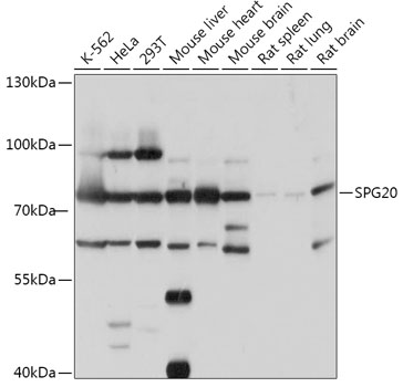
Figure 1. Western blot analysis of SPART using anti-SPART antibody (A31948-2). Electrophoresis was performed on a 5-20% SDS-PAGE gel at 70V (Stacking gel) / 90V (Resolving gel) for 2-3 hours. The sample well of each lane was loaded with 30 ug of sample under reducing conditions. Lane 1: human HEK293 whole cell lysates, Lane 2: human Hela whole cell lysates, Lane 3: rat brain tissue lysates, Lane 4: rat C6 whole cell lysates. After electrophoresis, proteins were transferred to a nitrocellulose membrane at 150 mA for 50-90 minutes. Blocked the membrane with 5% non-fat milk/TBS for 1.5 hour at RT. The membrane was incubated with rabbit anti-SPART antigen affinity purified polyclonal antibody (Catalog # A31948-2) at 0.5 microg/mL overnight at 4°C, then washed with TBS-0.1%Tween 3 times with 5 minutes each and probed with a goat anti-rabbit IgG-HRP secondary antibody at a dilution of 1:5000 for 1.5 hour at RT. The signal is developed using an Enhanced Chemiluminescent detection (ECL) kit (Catalog # EK1002) with Tanon 5200 system. A specific band was detected for SPART at approximately 78 kDa. The expected band size for SPART is at 73 kDa.
. Overlay histogram showing THP-1 cells stained with A31948-2 (Blue line). To facilitate intracellular staining, cells were fixed with 4% paraformaldehyde and permeabilized with permeabilization buffer. The cells were blocked with 10% normal goat serum. And then incubated with rabbit anti-SPART Antibody (A31948-2, 1 microg/1x106 cells) for 30 min at 20°C. DyLight®488 conjugated goat anti-rabbit IgG (BA1127, 5-10 microg/1x106 cells) was used as secondary antibody for 30 minutes at 20°C. Isotype control antibody (Green line) was rabbit IgG (1 microg/1x106) used under the same conditions. Unlabelled sample without incubation with primary antibody and secondary antibody (Red line) was used as a blank control.")
Figure 1. Western blot analysis of SPART using anti-SPART antibody (A31948-2). Electrophoresis was performed on a 5-20% SDS-PAGE gel at 70V (Stacking gel) / 90V (Resolving gel) for 2-3 hours. The sample well of each lane was loaded with 30 ug of sample under reducing conditions. Lane 1: human HEK293 whole cell lysates, Lane 2: human Hela whole cell lysates, Lane 3: rat brain tissue lysates, Lane 4: rat C6 whole cell lysates. After electrophoresis, proteins were transferred to a nitrocellulose membrane at 150 mA for 50-90 minutes. Blocked the membrane with 5% non-fat milk/TBS for 1.5 hour at RT. The membrane was incubated with rabbit anti-SPART antigen affinity purified polyclonal antibody (Catalog # A31948-2) at 0.5 microg/mL overnight at 4°C, then washed with TBS-0.1%Tween 3 times with 5 minutes each and probed with a goat anti-rabbit IgG-HRP secondary antibody at a dilution of 1:5000 for 1.5 hour at RT. The signal is developed using an Enhanced Chemiluminescent detection (ECL) kit (Catalog # EK1002) with Tanon 5200 system. A specific band was detected for SPART at approximately 78 kDa. The expected band size for SPART is at 73 kDa.
Anti-SPART Antibody Picoband(r)
A31948-2-CARRIER-FREE
ApplicationsFlow Cytometry, Western Blot, ELISA
Product group Antibodies
ReactivityHuman, Rat
TargetSPART
Overview
- SupplierBoster Bio
- Product NameAnti-SPART Antibody Picoband(r)
- Delivery Days Customer9
- ApplicationsFlow Cytometry, Western Blot, ELISA
- CertificationResearch Use Only
- ClonalityPolyclonal
- Concentration500 ug/ml
- Gene ID23111
- Target nameSPART
- Target descriptionspartin
- Target synonymsSPG20, TAHCCP1, spartin, spastic paraplegia 20 (Troyer syndrome), trans-activated by hepatitis C virus core protein 1
- HostRabbit
- IsotypeIgG
- Protein IDQ8N0X7
- Protein NameSpartin
- Scientific DescriptionBoster Bio Anti-SPART Antibody Picoband® catalog # A31948-2. Tested in ELISA, Flow Cytometry, WB applications. This antibody reacts with Human, Rat. The brand Picoband indicates this is a premium antibody that guarantees superior quality, high affinity, and strong signals with minimal background in Western blot applications. Only our best-performing antibodies are designated as Picoband, ensuring unmatched performance.
- ReactivityHuman, Rat
- Storage Instruction-20°C,2°C to 8°C
- UNSPSC12352203
Related products
Product group Antibodies
Anti-SPART AntibodyA307268
ApplicationsWestern Blot
ReactivityHuman, Mouse, Rat
- SizePrice
Product group Antibodies
Anti-SPG20 (C-term) Antibody102-22244
ApplicationsWestern Blot
TargetSPART
- SizePrice
Product group Antibodies

SPARTIN AntibodyABX433315
ApplicationsImmunoFluorescence, Western Blot, ELISA, ImmunoCytoChemistry
- SizePrice
Product group Antibodies

SPG20 / SPARTIN Antibody (aa500-550)LS-C762890
ApplicationsImmunoPrecipitation
ReactivityHuman
TargetSPART
- SizePrice
Product group Antibodies

Goat anti-SPARTINEB09136
ApplicationsImmunoFluorescence, Western Blot, ELISA
ReactivityHuman
TargetSPART
- SizePrice
Product group Antibodies
SPG20 AntibodyCSB-PA022542GA01HU
ApplicationsImmunoFluorescence, Western Blot, ELISA, ImmunoHistoChemistry
ReactivityHuman, Mouse, Rat
TargetSPART
- SizePrice
Product group Antibodies
SPARTIN antibody, InternalGTX88225
ApplicationsImmunoFluorescence, Western Blot, ImmunoCytoChemistry
ReactivityHuman
TargetSPART
- SizePrice

Product group Antibodies
Anti-SPG20 AntibodyHPA039053
ApplicationsImmunoHistoChemistry
ReactivityHuman
TargetSPART
- SizePrice