


Immunohistochemical staining of human urinary bladder shows strong cytoplasmic positivity in urothelial cells.

Immunohistochemical staining of human urinary bladder shows strong cytoplasmic positivity in urothelial cells.
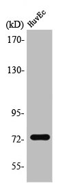
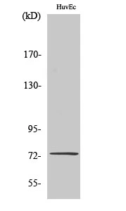
Anti-SSH3 Antibody
HPA019949
ApplicationsImmunoCytoChemistry, ImmunoHistoChemistry
Product group Antibodies
ReactivityHuman
TargetSSH3
Overview
- SupplierAtlas Antibodies
- Product NameAnti-SSH3 Antibody
- Delivery Days Customer4
- ApplicationsImmunoCytoChemistry, ImmunoHistoChemistry
- CertificationResearch Use Only
- ClonalityPolyclonal
- ConjugateUnconjugated
- Gene ID54961
- Target nameSSH3
- Target descriptionslingshot protein phosphatase 3
- Target synonymsSSH3L, protein phosphatase Slingshot homolog 3, SSH-3L, SSH-like protein 3, hSSH-3L, slingshot 3, slingshot homolog 3
- HostRabbit
- IsotypeIgG
- Protein IDQ8TE77
- Protein NameProtein phosphatase Slingshot homolog 3
- Scientific DescriptionRecombinant Protein Epitope Signature Tag (PrEST) antigen sequence
- ReactivityHuman
- Storage Instruction-20°C,2°C to 8°C
- UNSPSC41116161
Datasheet
MSDS
Related products
Product group Antibodies
SSH3 AntibodyCSB-PA004163
ApplicationsWestern Blot, ELISA, ImmunoHistoChemistry
ReactivityHuman, Mouse, Rat
TargetSSH3
- SizePrice
Product group Antibodies
SSH3 Polyclonal AntibodyCAC15325
ApplicationsImmunoFluorescence, Western Blot, ELISA, ImmunoHistoChemistry
ReactivityRat
TargetSSH3
- SizePrice

Product group Antibodies
Anti-SSH3 Antibody Picoband(r)A11609-3-CARRIER-FREE
ApplicationsFlow Cytometry, ImmunoFluorescence, Western Blot, ELISA, ImmunoCytoChemistry, ImmunoHistoChemistry
ReactivityHuman, Rat
TargetSSH3
- SizePrice

Product group Antibodies
Anti-SSH3 AntibodyA96120
ApplicationsWestern Blot, ELISA, ImmunoHistoChemistry
ReactivityHuman, Mouse, Rat
- SizePrice
Product group Antibodies

SSH3 AntibodyLS-C830990
ApplicationsELISA, ImmunoHistoChemistry
ReactivityHuman
TargetSSH3
- SizePrice

Product group Antibodies
Anti-SSH3 AntibodyHPA019957
ApplicationsImmunoHistoChemistry
ReactivityHuman
TargetSSH3
- SizePrice
Product group Antibodies
SSH3 antibodyGTX34228
ApplicationsWestern Blot
ReactivityHuman
TargetSSH3
- SizePrice
Product group Antibodies
Anti-SSH3 (Ser37) Antibody102-25260
ApplicationsWestern Blot
TargetSSH3
- SizePrice