


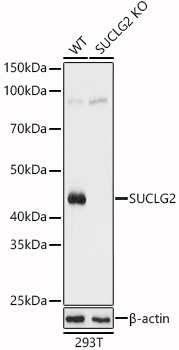
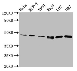
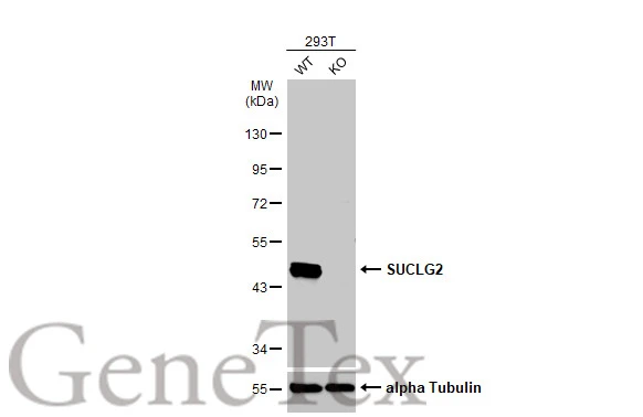
Figure 1. Western blot analysis of SUCLG2 using anti-SUCLG2 antibody (A08268-1). Electrophoresis was performed on a 5-20% SDS-PAGE gel at 70V (Stacking gel) / 90V (Resolving gel) for 2-3 hours. The sample well of each lane was loaded with 30 ug of sample under reducing conditions. Lane 1: human 293T whole cell lysates, Lane 2: human Hela whole cell lysates, Lane 3: human HepG2 whole cell lysates, Lane 4: human COLO320 whole cell lysates, Lane 5: rat liver tissue lysates, Lane 6: mouse liver tissue lysates. After electrophoresis, proteins were transferred to a nitrocellulose membrane at 150 mA for 50-90 minutes. Blocked the membrane with 5% non-fat milk/TBS for 1.5 hour at RT. The membrane was incubated with rabbit anti-SUCLG2 antigen affinity purified polyclonal antibody (Catalog # A08268-1) at 0.5 microg/mL overnight at 4°C, then washed with TBS-0.1%Tween 3 times with 5 minutes each and probed with a goat anti-rabbit IgG-HRP secondary antibody at a dilution of 1:5000 for 1.5 hour at RT. The signal is developed using an Enhanced Chemiluminescent detection (ECL) kit (Catalog # EK1002) with Tanon 5200 system. A specific band was detected for SUCLG2 at approximately 47 kDa. The expected band size for SUCLG2 is at 47 kDa.
. SUCLG2 was detected in a paraffin-embedded section of human breast cancer tissue. Heat mediated antigen retrieval was performed in EDTA buffer (pH 8.0, epitope retrieval solution). The tissue section was blocked with 10% goat serum. The tissue section was then incubated with 2 microg/ml rabbit anti-SUCLG2 Antibody (A08268-1) overnight at 4°C. Peroxidase Conjugated Goat Anti-rabbit IgG was used as secondary antibody and incubated for 30 minutes at 37°C. The tissue section was developed using HRP Conjugated Rabbit IgG Super Vision Assay Kit (Catalog # SV0002) with DAB as the chromogen.")
. SUCLG2 was detected in a paraffin-embedded section of human liver cancer tissue. Heat mediated antigen retrieval was performed in EDTA buffer (pH 8.0, epitope retrieval solution). The tissue section was blocked with 10% goat serum. The tissue section was then incubated with 2 microg/ml rabbit anti-SUCLG2 Antibody (A08268-1) overnight at 4°C. Peroxidase Conjugated Goat Anti-rabbit IgG was used as secondary antibody and incubated for 30 minutes at 37°C. The tissue section was developed using HRP Conjugated Rabbit IgG Super Vision Assay Kit (Catalog # SV0002) with DAB as the chromogen.")
. SUCLG2 was detected in a paraffin-embedded section of human ovarian serous adenocarcinoma tissue. Heat mediated antigen retrieval was performed in EDTA buffer (pH 8.0, epitope retrieval solution). The tissue section was blocked with 10% goat serum. The tissue section was then incubated with 2 microg/ml rabbit anti-SUCLG2 Antibody (A08268-1) overnight at 4°C. Peroxidase Conjugated Goat Anti-rabbit IgG was used as secondary antibody and incubated for 30 minutes at 37°C. The tissue section was developed using HRP Conjugated Rabbit IgG Super Vision Assay Kit (Catalog # SV0002) with DAB as the chromogen.")
. SUCLG2 was detected in a paraffin-embedded section of human prostate adenocarcinoma tissue. Heat mediated antigen retrieval was performed in EDTA buffer (pH 8.0, epitope retrieval solution). The tissue section was blocked with 10% goat serum. The tissue section was then incubated with 2 microg/ml rabbit anti-SUCLG2 Antibody (A08268-1) overnight at 4°C. Peroxidase Conjugated Goat Anti-rabbit IgG was used as secondary antibody and incubated for 30 minutes at 37°C. The tissue section was developed using HRP Conjugated Rabbit IgG Super Vision Assay Kit (Catalog # SV0002) with DAB as the chromogen.")
. SUCLG2 was detected in a paraffin-embedded section of human rectum adenocarcinoma tissue. Heat mediated antigen retrieval was performed in EDTA buffer (pH 8.0, epitope retrieval solution). The tissue section was blocked with 10% goat serum. The tissue section was then incubated with 2 microg/ml rabbit anti-SUCLG2 Antibody (A08268-1) overnight at 4°C. Peroxidase Conjugated Goat Anti-rabbit IgG was used as secondary antibody and incubated for 30 minutes at 37°C. The tissue section was developed using HRP Conjugated Rabbit IgG Super Vision Assay Kit (Catalog # SV0002) with DAB as the chromogen.")
. SUCLG2 was detected in a paraffin-embedded section of human spleen tissue. Heat mediated antigen retrieval was performed in EDTA buffer (pH 8.0, epitope retrieval solution). The tissue section was blocked with 10% goat serum. The tissue section was then incubated with 2 microg/ml rabbit anti-SUCLG2 Antibody (A08268-1) overnight at 4°C. Peroxidase Conjugated Goat Anti-rabbit IgG was used as secondary antibody and incubated for 30 minutes at 37°C. The tissue section was developed using HRP Conjugated Rabbit IgG Super Vision Assay Kit (Catalog # SV0002) with DAB as the chromogen.")
. SUCLG2 was detected in a paraffin-embedded section of human lung adenocarcinoma tissue. Heat mediated antigen retrieval was performed in EDTA buffer (pH 8.0, epitope retrieval solution). The tissue section was blocked with 10% goat serum. The tissue section was then incubated with 2 microg/ml rabbit anti-SUCLG2 Antibody (A08268-1) overnight at 4°C. Peroxidase Conjugated Goat Anti-rabbit IgG was used as secondary antibody and incubated for 30 minutes at 37°C. The tissue section was developed using HRP Conjugated Rabbit IgG Super Vision Assay Kit (Catalog # SV0002) with DAB as the chromogen.")
. SUCLG2 was detected in a paraffin-embedded section of human thyroid papillary carcinoma tissue. Heat mediated antigen retrieval was performed in EDTA buffer (pH 8.0, epitope retrieval solution). The tissue section was blocked with 10% goat serum. The tissue section was then incubated with 2 microg/ml rabbit anti-SUCLG2 Antibody (A08268-1) overnight at 4°C. Peroxidase Conjugated Goat Anti-rabbit IgG was used as secondary antibody and incubated for 30 minutes at 37°C. The tissue section was developed using HRP Conjugated Rabbit IgG Super Vision Assay Kit (Catalog # SV0002) with DAB as the chromogen.")
. SUCLG2 was detected in a paraffin-embedded section of mouse kidney tissue. Heat mediated antigen retrieval was performed in EDTA buffer (pH 8.0, epitope retrieval solution). The tissue section was blocked with 10% goat serum. The tissue section was then incubated with 2 microg/ml rabbit anti-SUCLG2 Antibody (A08268-1) overnight at 4°C. Peroxidase Conjugated Goat Anti-rabbit IgG was used as secondary antibody and incubated for 30 minutes at 37°C. The tissue section was developed using HRP Conjugated Rabbit IgG Super Vision Assay Kit (Catalog # SV0002) with DAB as the chromogen.")
Figure 1. Western blot analysis of SUCLG2 using anti-SUCLG2 antibody (A08268-1). Electrophoresis was performed on a 5-20% SDS-PAGE gel at 70V (Stacking gel) / 90V (Resolving gel) for 2-3 hours. The sample well of each lane was loaded with 30 ug of sample under reducing conditions. Lane 1: human 293T whole cell lysates, Lane 2: human Hela whole cell lysates, Lane 3: human HepG2 whole cell lysates, Lane 4: human COLO320 whole cell lysates, Lane 5: rat liver tissue lysates, Lane 6: mouse liver tissue lysates. After electrophoresis, proteins were transferred to a nitrocellulose membrane at 150 mA for 50-90 minutes. Blocked the membrane with 5% non-fat milk/TBS for 1.5 hour at RT. The membrane was incubated with rabbit anti-SUCLG2 antigen affinity purified polyclonal antibody (Catalog # A08268-1) at 0.5 microg/mL overnight at 4°C, then washed with TBS-0.1%Tween 3 times with 5 minutes each and probed with a goat anti-rabbit IgG-HRP secondary antibody at a dilution of 1:5000 for 1.5 hour at RT. The signal is developed using an Enhanced Chemiluminescent detection (ECL) kit (Catalog # EK1002) with Tanon 5200 system. A specific band was detected for SUCLG2 at approximately 47 kDa. The expected band size for SUCLG2 is at 47 kDa.
Anti-SUCLG2 Antibody Picoband(r)
A08268-1-CARRIER-FREE
ApplicationsFlow Cytometry, ImmunoFluorescence, Western Blot, ELISA, ImmunoCytoChemistry, ImmunoHistoChemistry
Product group Antibodies
ReactivityHuman, Mouse, Rat
TargetSUCLG2
Overview
- SupplierBoster Bio
- Product NameAnti-SUCLG2 Antibody Picoband(r)
- Delivery Days Customer9
- ApplicationsFlow Cytometry, ImmunoFluorescence, Western Blot, ELISA, ImmunoCytoChemistry, ImmunoHistoChemistry
- CertificationResearch Use Only
- ClonalityPolyclonal
- Concentration500 ug/ml
- Gene ID8801
- Target nameSUCLG2
- Target descriptionsuccinate-CoA ligase GDP-forming subunit beta
- Target synonymsG-SCS, GBETA, GTPSCS, succinate--CoA ligase [GDP-forming] subunit beta, mitochondrial, GTP-specific succinyl-CoA synthetase beta subunit, GTP-specific succinyl-CoA synthetase subunit beta, SCS-betaG, succinate-CoA ligase GDP-forming beta subunit, succinyl-CoA ligase [GDP-forming] subunit beta, mitochondrial, succinyl-CoA ligase, GDP-forming, beta chain, mitochondrial, succinyl-CoA synthetase, beta-G chain
- HostRabbit
- IsotypeIgG
- Protein IDQ96I99
- Protein NameSuccinate--CoA ligase [GDP-forming] subunit beta, mitochondrial
- Scientific DescriptionBoster Bio Anti-SUCLG2 Antibody Picoband® catalog # A08268-1. Tested in ELISA, Flow Cytometry, IF, IHC, ICC, WB applications. This antibody reacts with Human, Mouse, Rat. The brand Picoband indicates this is a premium antibody that guarantees superior quality, high affinity, and strong signals with minimal background in Western blot applications. Only our best-performing antibodies are designated as Picoband, ensuring unmatched performance.
- ReactivityHuman, Mouse, Rat
- Storage Instruction-20°C,2°C to 8°C
- UNSPSC12352203
Related products
Product group Antibodies
Anti-SUCLG2 AntibodyA90091
ApplicationsImmunoFluorescence, Western Blot, ImmunoCytoChemistry, ImmunoHistoChemistry
ReactivityHuman, Mouse, Rat
- SizePrice

Product group Antibodies
Anti-SUCLG2 AntibodyHPA046705
ApplicationsWestern Blot, ImmunoCytoChemistry, ImmunoHistoChemistry
ReactivityHuman
TargetSUCLG2
- SizePrice
Product group Antibodies

SUCLG2 AntibodyLS-C410510
ApplicationsWestern Blot, ImmunoHistoChemistry
ReactivityHuman, Mouse, Rat
TargetSUCLG2
- SizePrice
Product group Antibodies
SUCLG2 AntibodyCSB-PA846636LA01HU
ApplicationsWestern Blot, ELISA, ImmunoHistoChemistry
ReactivityHuman
TargetSUCLG2
- SizePrice
Product group Antibodies
SUCLG2 Polyclonal AntibodyCAC15384
ApplicationsWestern Blot, ELISA, ImmunoHistoChemistry
TargetSUCLG2
- SizePrice
Product group Antibodies
SUCLG2 antibodyGTX107002
ApplicationsImmunoFluorescence, Western Blot, ImmunoCytoChemistry, ImmunoHistoChemistry, ImmunoHistoChemistry Paraffin
ReactivityHuman, Mouse
TargetSUCLG2
- SizePrice
Product group Antibodies
Anti-SUCLG2 Antibody144-61461
ApplicationsWestern Blot
ReactivityHuman, Mouse, Rat
TargetSUCLG2
- SizePrice