

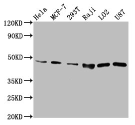
Western Blot Positive WB detected in: Hela whole cell lysate, MCF-7 whole cell lysate, 293T whole cell lysate, Raji whole cell lysate, LO2 whole cell lysate, U87 whole cell lysate, All lanes: SUCLG2 antibody at 6.7microg/ml Secondary Goat polyclonal to rabbit IgG at 1/50000 dilution Predicted band size: 47, 48 kDa Observed band size: 47 kDa
. Section was blocked with 10% normal goat serum 30min at RT. Then primary antibody (1% BSA) was incubated at 4°C overnight. The primary is detected by a biotinylated secondary antibody and visualized using an HRP conjugated SP system.")
. Section was blocked with 10% normal goat serum 30min at RT. Then primary antibody (1% BSA) was incubated at 4°C overnight. The primary is detected by a biotinylated secondary antibody and visualized using an HRP conjugated SP system.")
Western Blot Positive WB detected in: Hela whole cell lysate, MCF-7 whole cell lysate, 293T whole cell lysate, Raji whole cell lysate, LO2 whole cell lysate, U87 whole cell lysate, All lanes: SUCLG2 antibody at 6.7microg/ml Secondary Goat polyclonal to rabbit IgG at 1/50000 dilution Predicted band size: 47, 48 kDa Observed band size: 47 kDa
SUCLG2 Antibody
CSB-PA846636LA01HU
ApplicationsWestern Blot, ELISA, ImmunoHistoChemistry
Product group Antibodies
ReactivityHuman
TargetSUCLG2
Overview
- SupplierCusabio
- Product NameSUCLG2 Antibody
- Delivery Days Customer20
- ApplicationsWestern Blot, ELISA, ImmunoHistoChemistry
- CertificationResearch Use Only
- ClonalityPolyclonal
- ConjugateUnconjugated
- Gene ID8801
- Target nameSUCLG2
- Target descriptionsuccinate-CoA ligase GDP-forming subunit beta
- Target synonymsG-SCS, GBETA, GTPSCS, succinate--CoA ligase [GDP-forming] subunit beta, mitochondrial, GTP-specific succinyl-CoA synthetase beta subunit, GTP-specific succinyl-CoA synthetase subunit beta, SCS-betaG, succinate-CoA ligase GDP-forming beta subunit, succinyl-CoA ligase [GDP-forming] subunit beta, mitochondrial, succinyl-CoA ligase, GDP-forming, beta chain, mitochondrial, succinyl-CoA synthetase, beta-G chain
- HostRabbit
- IsotypeIgG
- Protein IDQ96I99
- Protein NameSuccinate--CoA ligase [GDP-forming] subunit beta, mitochondrial
- Scientific DescriptionGTP-specific succinyl-CoA synthetase functions in the citric acid cycle (TCA), coupling the hydrolysis of succinyl-CoA to the synthesis of GTP and thus represents the only step of substrate-level phosphorylation in the TCA. The beta subunit provides nucleotide specificity of the enzyme and binds the substrate succinate, while the binding sites for coenzyme A and phosphate are found in the alpha subunit.
- ReactivityHuman
- Storage Instruction-20°C or -80°C
- UNSPSC41116161
Related products

Product group Antibodies
Anti-SUCLG2 Antibody Picoband(r)A08268-1-CARRIER-FREE
ApplicationsFlow Cytometry, ImmunoFluorescence, Western Blot, ELISA, ImmunoCytoChemistry, ImmunoHistoChemistry
ReactivityHuman, Mouse, Rat
TargetSUCLG2
- SizePrice
Product group Antibodies
Anti-SUCLG2 AntibodyA90091
ApplicationsImmunoFluorescence, Western Blot, ImmunoCytoChemistry, ImmunoHistoChemistry
ReactivityHuman, Mouse, Rat
- SizePrice

Product group Antibodies
Anti-SUCLG2 AntibodyHPA046705
ApplicationsWestern Blot, ImmunoCytoChemistry, ImmunoHistoChemistry
ReactivityHuman
TargetSUCLG2
- SizePrice
Product group Antibodies

SUCLG2 AntibodyLS-C410510
ApplicationsWestern Blot, ImmunoHistoChemistry
ReactivityHuman, Mouse, Rat
TargetSUCLG2
- SizePrice
Product group Antibodies
SUCLG2 Polyclonal AntibodyCAC15384
ApplicationsWestern Blot, ELISA, ImmunoHistoChemistry
TargetSUCLG2
- SizePrice
Product group Antibodies
SUCLG2 antibodyGTX107002
ApplicationsImmunoFluorescence, Western Blot, ImmunoCytoChemistry, ImmunoHistoChemistry, ImmunoHistoChemistry Paraffin
ReactivityHuman, Mouse
TargetSUCLG2
- SizePrice
Product group Antibodies
Anti-SUCLG2 Antibody144-61461
ApplicationsWestern Blot
ReactivityHuman, Mouse, Rat
TargetSUCLG2
- SizePrice