


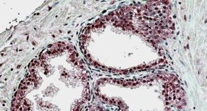
Immunohistochemical staining of human Testis shows strong nuclear positivity in cells in seminiferous ducts.

Immunohistochemical staining of human Testis shows strong nuclear positivity in cells in seminiferous ducts.
Anti-SUFU Antibody
HPA008700
ApplicationsWestern Blot, ImmunoCytoChemistry, ImmunoHistoChemistry
Product group Antibodies
ReactivityHuman
TargetSUFU
Overview
- SupplierAtlas Antibodies
- Product NameAnti-SUFU Antibody
- Delivery Days Customer4
- ApplicationsWestern Blot, ImmunoCytoChemistry, ImmunoHistoChemistry
- CertificationResearch Use Only
- ClonalityPolyclonal
- ConjugateUnconjugated
- Gene ID51684
- Target nameSUFU
- Target descriptionSUFU negative regulator of hedgehog signaling
- Target synonymsBCNS2, JBTS32, PRO1280, SUFUH, SUFUXL, suppressor of fused homolog, negative regulator of hedgehog signaling
- HostRabbit
- IsotypeIgG
- Protein IDQ9UMX1
- Protein NameSuppressor of fused homolog
- Scientific DescriptionRecombinant Protein Epitope Signature Tag (PrEST) antigen sequence
- ReactivityHuman
- Storage Instruction-20°C,2°C to 8°C
- UNSPSC41116161
Datasheet
MSDS
Related products
Product group Antibodies
Anti-SUFU Antibody144-06757
ApplicationsImmunoFluorescence, Western Blot, ImmunoHistoChemistry
ReactivityHuman, Mouse, Rat
TargetSUFU
- SizePrice
Product group Antibodies
Anti-SUFU AntibodyA326304
ApplicationsELISA, ImmunoHistoChemistry
ReactivityHuman
- SizePrice
Product group Antibodies

SUFU AntibodyLS-C748477
ApplicationsImmunoFluorescence, Western Blot
ReactivityHuman, Mouse, Rat
TargetSUFU
- SizePrice

Product group Antibodies
Anti-SUFU Picoband(r) AntibodyA02279-1-CARRIER-FREE
ApplicationsFlow Cytometry, ImmunoFluorescence, Western Blot, ELISA, ImmunoCytoChemistry
ReactivityHuman, Mouse, Rat
TargetSUFU
- SizePrice
Product group Antibodies
References
SUFU Polyclonal AntibodyBS-11209R
ApplicationsImmunoFluorescence, Western Blot, ELISA, ImmunoCytoChemistry, ImmunoHistoChemistry, ImmunoHistoChemistry Frozen, ImmunoHistoChemistry Paraffin
TargetSUFU
- SizePrice
Product group Antibodies
SUFU AntibodyCSB-PA891560ESR2HU
ApplicationsWestern Blot, ELISA, ImmunoHistoChemistry
ReactivityHuman, Mouse
TargetSUFU
- SizePrice
![Rat whole cell extracts (30 μg) was separated by 10% SDS-PAGE, and the membrane was blotted with Suppressor of Fused antibody [HL3374] (GTX641194) diluted at 1:1000. The HRP-conjugated anti-rabbit IgG antibody (GTX213110-01) was used to detect the primary antibody.](https://www.genetex.com/upload/website/prouct_img/normal/GTX641194/GTX641194_T-45572_20241025_WB_R_24103022_629.webp)
Product group Antibodies
ApplicationsWestern Blot, ImmunoHistoChemistry, ImmunoHistoChemistry Paraffin
ReactivityHuman, Mouse, Rat
TargetSUFU
- SizePrice
Product group Antibodies
Anti-SUFU AntibodyCAB13429
ApplicationsImmunoFluorescence, Western Blot, ELISA, ImmunoCytoChemistry, ImmunoHistoChemistry, ImmunoHistoChemistry Paraffin
ReactivityHuman
TargetSUFU
- SizePrice