


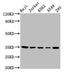
Figure 1. Western blot analysis of TAL1 using anti-TAL1 antibody (A00944-4). Electrophoresis was performed on a 5-20% SDS-PAGE gel at 70V (Stacking gel) / 90V (Resolving gel) for 2-3 hours. The sample well of each lane was loaded with 30 ug of sample under reducing conditions. Lane 1: human Jurkat whole cell lysates, Lane 2: human HEL whole cell lysates, Lane 3: human MOLT-4 whole cell lysates, Lane 4: rat lung tissue lysate, Lane 5: rat spleen tissue lysate, Lane 6: mouse lung tissue lysate, Lane 7: mouse spleen tissue lysate. After electrophoresis, proteins were transferred to a nitrocellulose membrane at 150 mA for 50-90 minutes. Blocked the membrane with 5% non-fat milk/TBS for 1.5 hour at RT. The membrane was incubated with rabbit anti-TAL1 antigen affinity purified polyclonal antibody (Catalog # A00944-4) at 0.25 microg/mL overnight at 4°C, then washed with TBS-0.1%Tween 3 times with 5 minutes each and probed with a goat anti-rabbit IgG-HRP secondary antibody at a dilution of 1:5000 for 1.5 hour at RT. The signal is developed using an Enhanced Chemiluminescent detection (ECL) kit (Catalog # EK1002) with Tanon 5200 system. A specific band was detected for TAL1 at approximately 40-45 kDa. The expected band size for TAL1 is at 34 kDa.
. Overlay histogram showing HEL cells stained with A00944-4 (Blue line). To facilitate intracellular staining, cells were fixed with 4% paraformaldehyde and permeabilized with permeabilization buffer. The cells were blocked with 10% normal goat serum. And then incubated with rabbit anti-TAL1 Antibody (A00944-4, 1 microg/1x106 cells) for 30 min at 20°C. DyLight®488 conjugated goat anti-rabbit IgG (BA1127, 5-10 microg/1x106 cells) was used as secondary antibody for 30 minutes at 20°C. Isotype control antibody (Green line) was rabbit IgG (1 microg/1x106) used under the same conditions. Unlabelled sample without incubation with primary antibody and secondary antibody (Red line) was used as a blank control.")
Figure 1. Western blot analysis of TAL1 using anti-TAL1 antibody (A00944-4). Electrophoresis was performed on a 5-20% SDS-PAGE gel at 70V (Stacking gel) / 90V (Resolving gel) for 2-3 hours. The sample well of each lane was loaded with 30 ug of sample under reducing conditions. Lane 1: human Jurkat whole cell lysates, Lane 2: human HEL whole cell lysates, Lane 3: human MOLT-4 whole cell lysates, Lane 4: rat lung tissue lysate, Lane 5: rat spleen tissue lysate, Lane 6: mouse lung tissue lysate, Lane 7: mouse spleen tissue lysate. After electrophoresis, proteins were transferred to a nitrocellulose membrane at 150 mA for 50-90 minutes. Blocked the membrane with 5% non-fat milk/TBS for 1.5 hour at RT. The membrane was incubated with rabbit anti-TAL1 antigen affinity purified polyclonal antibody (Catalog # A00944-4) at 0.25 microg/mL overnight at 4°C, then washed with TBS-0.1%Tween 3 times with 5 minutes each and probed with a goat anti-rabbit IgG-HRP secondary antibody at a dilution of 1:5000 for 1.5 hour at RT. The signal is developed using an Enhanced Chemiluminescent detection (ECL) kit (Catalog # EK1002) with Tanon 5200 system. A specific band was detected for TAL1 at approximately 40-45 kDa. The expected band size for TAL1 is at 34 kDa.
Anti-TAL1 Antibody Picoband(r)
A00944-4-IFLUOR647
ApplicationsFlow Cytometry, Western Blot
Product group Antibodies
ReactivityHuman, Mouse, Rat
TargetTAL1
Overview
- SupplierBoster Bio
- Product NameAnti-TAL1 Antibody Picoband(r)
- Delivery Days Customer9
- ApplicationsFlow Cytometry, Western Blot
- CertificationResearch Use Only
- ClonalityPolyclonal
- Concentration500 ug/ml
- ConjugateOther Conjugate
- Gene ID6886
- Target nameTAL1
- Target descriptionTAL bHLH transcription factor 1, erythroid differentiation factor
- Target synonymsSCL, TCL5, bHLHa17, tal-1, T-cell acute lymphocytic leukemia protein 1, T-cell acute lymphocytic leukemia 1, T-cell leukemia/lymphoma protein 5, class A basic helix-loop-helix protein 17, stem cell protein, tal-1 product
- HostRabbit
- IsotypeIgG
- Protein IDP17542
- Protein NameT-cell acute lymphocytic leukemia protein 1
- Scientific DescriptionBoster Bio Anti-TAL1 Antibody Picoband® catalog # A00944-4. Tested in Flow Cytometry, WB applications. This antibody reacts with Human, Mouse, Rat. The brand Picoband indicates this is a premium antibody that guarantees superior quality, high affinity, and strong signals with minimal background in Western blot applications. Only our best-performing antibodies are designated as Picoband, ensuring unmatched performance.
- ReactivityHuman, Mouse, Rat
- Storage Instruction-20°C,2°C to 8°C
- UNSPSC12352203
Related products
Product group Antibodies
TAL1 Polyclonal AntibodyCAC15315
ApplicationsImmunoFluorescence, Western Blot, ELISA
TargetTAL1
- SizePrice
Product group Antibodies
ApplicationsWestern Blot
ReactivityHuman, Mouse
- SizePrice
Product group Antibodies
Anti-TAL1 Antibody144-12927
ApplicationsWestern Blot
ReactivityHuman, Rat
TargetTAL1
- SizePrice
Product group Antibodies

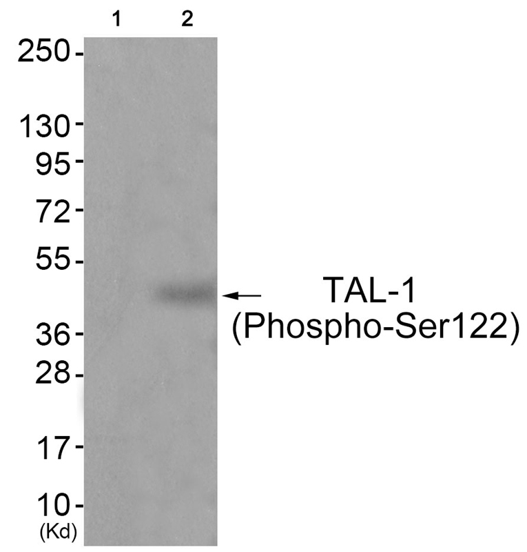
TAL-1 (Phospho-Ser122) AntibodyABX012709
ApplicationsWestern Blot, ELISA
- SizePrice
Product group Antibodies
References
TAL1 antibody [C2C3], C-termGTX116020
ApplicationsImmunoPrecipitation, Western Blot, ChIP Chromatin ImmunoPrecipitation, ImmunoHistoChemistry, ImmunoHistoChemistry Paraffin
ReactivityHuman, Mouse
TargetTAL1
- SizePrice
Product group Antibodies

TAL1 AntibodyLS-C747995
ApplicationsWestern Blot
ReactivityHuman, Rat
TargetTAL1
- SizePrice
Product group Antibodies
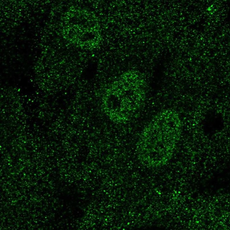
Anti-TAL1 AntibodyHPA073983
ApplicationsChIP Chromatin ImmunoPrecipitation, ImmunoCytoChemistry
ReactivityHuman
TargetTAL1
- SizePrice
Product group Antibodies
TAL1 AntibodyCSB-PA023110LA01HU
ApplicationsImmunoFluorescence, Western Blot, ELISA
ReactivityHuman
TargetTAL1
- SizePrice