


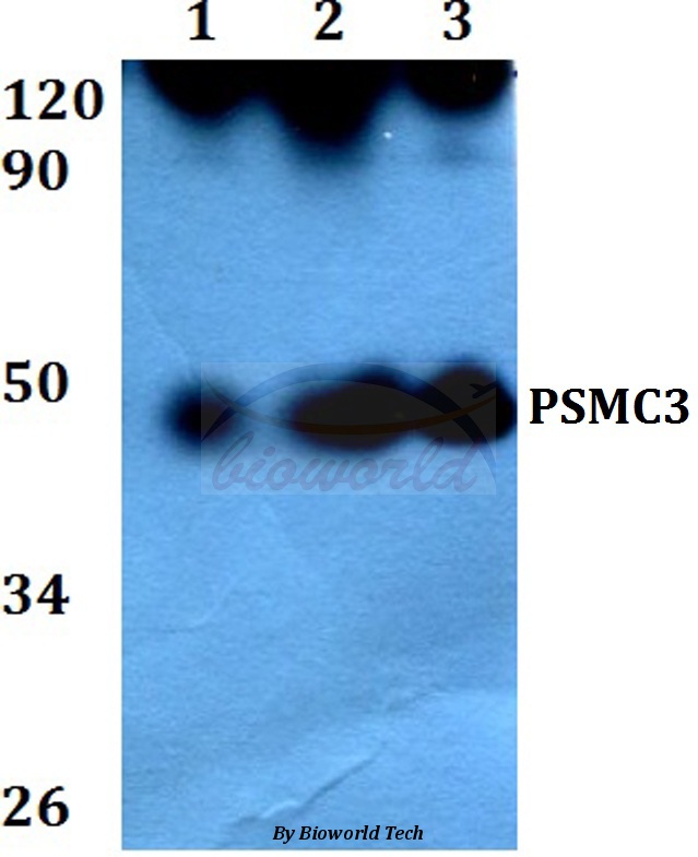
Figure 1. Western blot analysis of TBP-1/PSMC3 using anti-TBP-1/PSMC3 antibody (A07208-1). Electrophoresis was performed on a 5-20% SDS-PAGE gel at 70V (Stacking gel) / 90V (Resolving gel) for 2-3 hours. The sample well of each lane was loaded with 50ug of sample under reducing conditions. Lane 1: human HEK293 whole cell lysates, Lane 2: human HELA whole cell lysates, Lane 3: human HEPG2 whole cell lysates, Lane 4: human MCF-7 whole cell lysates, Lane 5: human U20S whole cell lysates, Lane 6: human PC-3 whole cell lysates, Lane 7: human U87 whole cell lysates, Lane 8: rat stomach tissue lysates, Lane 9: mouse lung tissue lysates. After Electrophoresis, proteins were transferred to a Nitrocellulose membrane at 150mA for 50-90 minutes. Blocked the membrane with 5% Non-fat Milk/ TBS for 1.5 hour at RT. The membrane was incubated with rabbit anti-TBP-1/PSMC3 antigen affinity purified polyclonal antibody (Catalog # A07208-1) at 0.25 microg/mL overnight at 4°C, then washed with TBS-0.1%Tween 3 times with 5 minutes each and probed with a goat anti-rabbit IgG-HRP secondary antibody at a dilution of 1:5000 for 1.5 hour at RT. The signal is developed using an Enhanced Chemiluminescent detection (ECL) kit (Catalog # EK1002) with Tanon 5200 system. A specific band was detected for TBP-1/PSMC3 at approximately 50KD. The expected band size for TBP-1/PSMC3 is at 50KD.
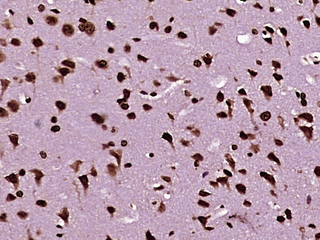
. TBP-1/PSMC3 was detected in paraffin-embedded section of rat cardiac muscle tissue. Heat mediated antigen retrieval was performed in EDTA buffer (pH8.0, epitope retrieval solution). The tissue section was blocked with 10% goat serum. The tissue section was then incubated with 2microg/ml rabbit anti-TBP-1/PSMC3 Antibody (A07208-1) overnight at 4°C. Biotinylated goat anti-rabbit IgG was used as secondary antibody and incubated for 30 minutes at 37°C. The tissue section was developed using Strepavidin-Biotin-Complex (SABC) (Catalog # SA1022) with DAB as the chromogen.")
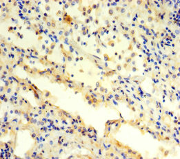
. TBP-1/PSMC3 was detected in paraffin-embedded section of human appendicitis tissue. Heat mediated antigen retrieval was performed in EDTA buffer (pH8.0, epitope retrieval solution). The tissue section was blocked with 10% goat serum. The tissue section was then incubated with 2microg/ml rabbit anti-TBP-1/PSMC3 Antibody (A07208-1) overnight at 4°C. Biotinylated goat anti-rabbit IgG was used as secondary antibody and incubated for 30 minutes at 37°C. The tissue section was developed using Strepavidin-Biotin-Complex (SABC) (Catalog # SA1022) with DAB as the chromogen.")
. TBP-1/PSMC3 was detected in paraffin-embedded section of human renal carcinoma tissue. Heat mediated antigen retrieval was performed in EDTA buffer (pH8.0, epitope retrieval solution). The tissue section was blocked with 10% goat serum. The tissue section was then incubated with 2microg/ml rabbit anti-TBP-1/PSMC3 Antibody (A07208-1) overnight at 4°C. Biotinylated goat anti-rabbit IgG was used as secondary antibody and incubated for 30 minutes at 37°C. The tissue section was developed using Strepavidin-Biotin-Complex (SABC) (Catalog # SA1022) with DAB as the chromogen.")
. TBP-1/PSMC3 was detected in paraffin-embedded section of human bladder tissue. Heat mediated antigen retrieval was performed in EDTA buffer (pH8.0, epitope retrieval solution). The tissue section was blocked with 10% goat serum. The tissue section was then incubated with 2microg/ml rabbit anti-TBP-1/PSMC3 Antibody (A07208-1) overnight at 4°C. Biotinylated goat anti-rabbit IgG was used as secondary antibody and incubated for 30 minutes at 37°C. The tissue section was developed using Strepavidin-Biotin-Complex (SABC) (Catalog # SA1022) with DAB as the chromogen.")
. TBP-1/PSMC3 was detected in paraffin-embedded section of human pancreatic cancer tissue. Heat mediated antigen retrieval was performed in EDTA buffer (pH8.0, epitope retrieval solution). The tissue section was blocked with 10% goat serum. The tissue section was then incubated with 2microg/ml rabbit anti-TBP-1/PSMC3 Antibody (A07208-1) overnight at 4°C. Biotinylated goat anti-rabbit IgG was used as secondary antibody and incubated for 30 minutes at 37°C. The tissue section was developed using Strepavidin-Biotin-Complex (SABC) (Catalog # SA1022) with DAB as the chromogen.")
. Overlay histogram showing 293T cells stained with A07208-1 (Blue line). To facilitate intracellular staining, cells were fixed with 4% paraformaldehyde and permeabilized with permeabilization buffer. The cells were blocked with 10% normal goat serum. And then incubated with rabbit anti-TBP-1/PSMC3 Antibody (A07208-1, 1microg/1x106 cells) for 30 min at 20°C. DyLight®488 conjugated goat anti-rabbit IgG (BA1127, 5-10microg/1x106 cells) was used as secondary antibody for 30 minutes at 20°C. Isotype control antibody (Green line) was rabbit IgG (1microg/1x106) used under the same conditions. Unlabelled sample without incubation with primary antibody and secondary antibody (Red line) was used as a blank control.")
Figure 1. Western blot analysis of TBP-1/PSMC3 using anti-TBP-1/PSMC3 antibody (A07208-1). Electrophoresis was performed on a 5-20% SDS-PAGE gel at 70V (Stacking gel) / 90V (Resolving gel) for 2-3 hours. The sample well of each lane was loaded with 50ug of sample under reducing conditions. Lane 1: human HEK293 whole cell lysates, Lane 2: human HELA whole cell lysates, Lane 3: human HEPG2 whole cell lysates, Lane 4: human MCF-7 whole cell lysates, Lane 5: human U20S whole cell lysates, Lane 6: human PC-3 whole cell lysates, Lane 7: human U87 whole cell lysates, Lane 8: rat stomach tissue lysates, Lane 9: mouse lung tissue lysates. After Electrophoresis, proteins were transferred to a Nitrocellulose membrane at 150mA for 50-90 minutes. Blocked the membrane with 5% Non-fat Milk/ TBS for 1.5 hour at RT. The membrane was incubated with rabbit anti-TBP-1/PSMC3 antigen affinity purified polyclonal antibody (Catalog # A07208-1) at 0.25 microg/mL overnight at 4°C, then washed with TBS-0.1%Tween 3 times with 5 minutes each and probed with a goat anti-rabbit IgG-HRP secondary antibody at a dilution of 1:5000 for 1.5 hour at RT. The signal is developed using an Enhanced Chemiluminescent detection (ECL) kit (Catalog # EK1002) with Tanon 5200 system. A specific band was detected for TBP-1/PSMC3 at approximately 50KD. The expected band size for TBP-1/PSMC3 is at 50KD.
Anti-TBP-1/PSMC3 Antibody Picoband(r)
A07208-1-CARRIER-FREE
ApplicationsFlow Cytometry, Western Blot, ELISA, ImmunoHistoChemistry
Product group Antibodies
ReactivityHuman, Mouse, Rat
TargetPSMC3
Overview
- SupplierBoster Bio
- Product NameAnti-TBP-1/PSMC3 Antibody Picoband(r)
- Delivery Days Customer9
- ApplicationsFlow Cytometry, Western Blot, ELISA, ImmunoHistoChemistry
- CertificationResearch Use Only
- ClonalityPolyclonal
- Concentration500 ug/ml
- Gene ID5702
- Target namePSMC3
- Target descriptionproteasome 26S subunit, ATPase 3
- Target synonymsDCIDP, RPT5, TBP1, 26S proteasome regulatory subunit 6A, 26S protease regulatory subunit 6A, 26S proteasome AAA-ATPase subunit RPT5, Tat-binding protein 1, human immunodeficiency virus tat transactivator binding protein-1, proteasome (prosome, macropain) 26S subunit, ATPase, 3, proteasome subunit P50, testicular secretory protein Li 42
- HostRabbit
- IsotypeIgG
- Protein IDP17980
- Protein Name26S proteasome regulatory subunit 6A
- Scientific DescriptionBoster Bio Anti-TBP-1/PSMC3 Antibody Picoband® catalog # A07208-1. Tested in ELISA, Flow Cytometry, IHC, WB applications. This antibody reacts with Human, Mouse, Rat. The brand Picoband indicates this is a premium antibody that guarantees superior quality, high affinity, and strong signals with minimal background in Western blot applications. Only our best-performing antibodies are designated as Picoband, ensuring unmatched performance.
- ReactivityHuman, Mouse, Rat
- Storage Instruction-20°C,2°C to 8°C
- UNSPSC12352203
Related products
Product group Antibodies
PSMC3 AntibodyCSB-PA018891LA01HU
ApplicationsImmunoFluorescence, ELISA, ImmunoHistoChemistry
ReactivityHuman
TargetPSMC3
- SizePrice
Product group Antibodies
ApplicationsImmunoPrecipitation, Western Blot, ImmunoCytoChemistry, ImmunoHistoChemistry
ReactivityMouse, Rat
TargetPSMC3
- SizePrice
Product group Antibodies
Anti-PSMC3 Antibody144-01986
ApplicationsImmunoFluorescence, Western Blot, ImmunoHistoChemistry
ReactivityHuman, Mouse, Rat
TargetPSMC3
- SizePrice
Product group Antibodies
ApplicationsWestern Blot, ImmunoHistoChemistry
ReactivityHuman, Mouse, Rat
- SizePrice

Product group Antibodies
Anti-PSMC3 AntibodyHPA006065
ApplicationsWestern Blot, ImmunoCytoChemistry, ImmunoHistoChemistry
ReactivityHuman, Mouse, Rat
TargetPSMC3
- SizePrice
Product group Antibodies

PSMC3 Antibody (Internal)LS-C368682
ApplicationsWestern Blot, ImmunoHistoChemistry, ImmunoHistoChemistry Paraffin
ReactivityHuman, Mouse, Rat, Zebra Fish
TargetPSMC3
- SizePrice
Product group Antibodies
PSMC3 Polyclonal AntibodyBS-19463R
ApplicationsImmunoFluorescence, Western Blot, ELISA, ImmunoCytoChemistry, ImmunoHistoChemistry, ImmunoHistoChemistry Frozen, ImmunoHistoChemistry Paraffin
ReactivityBovine, Canine, Equine, Human, Mouse, Xenopus
TargetPSMC3
- SizePrice
![Immunofluorescent image of a zebrafish embryo eye section using PSMC3 antibody [N1C2] (GTX109605) at a 1:200 dilution. Psmc3 (Green) Actin(Red)(This image was provided courtesy of the Schilling Lab at UC, Irvine.)](https://www.genetex.com/upload/website/prouct_img/normal/GTX109605/GTX109605_40023_IHC-Wm_Z_22111423_320.webp)
Product group Antibodies
PSMC3 antibody [N1C2]GTX109605
ApplicationsImmunoFluorescence, Western Blot, ImmunoCytoChemistry, ImmunoHistoChemistry, ImmunoHistoChemistry Paraffin
ReactivityHuman, Zebra Fish
TargetPSMC3
- SizePrice