

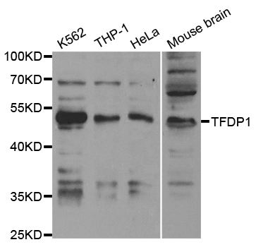
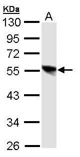
Anti-TFDP1 Antibody
144-63448
ApplicationsImmunoFluorescence, Western Blot, ImmunoHistoChemistry
Product group Antibodies
ReactivityHuman, Mouse, Rat
TargetTFDP1
Overview
- SupplierRayBiotech
- Product NameAnti-TFDP1 Antibody
- Delivery Days Customer16
- ApplicationsImmunoFluorescence, Western Blot, ImmunoHistoChemistry
- CertificationResearch Use Only
- ConjugateUnconjugated
- Gene ID7027
- Target nameTFDP1
- Target descriptiontranscription factor Dp-1
- Target synonymsDILC, DP1, DRTF1, Dp-1, transcription factor Dp-1, DRTF1-polypeptide 1, E2F dimerization partner 1, E2F-related transcription factor, down-regulated in liver cancer stem cells
- HostRabbit
- IsotypeIgG
- Protein IDQ14186
- Protein NameTranscription factor Dp-1
- Scientific DescriptionTFDP1 pAb
- ReactivityHuman, Mouse, Rat
- Storage Instruction-20°C
- UNSPSC12352203
Related products
Product group Antibodies
Anti-TFDP1 AntibodyA30466
ApplicationsWestern Blot, ImmunoHistoChemistry
ReactivityHuman, Mouse, Rat
- SizePrice
Product group Antibodies
ApplicationsFlow Cytometry, Western Blot
ReactivityHuman, Mouse, Rat
TargetTFDP1
- SizePrice

Product group Antibodies
TFDP1 AntibodyCSB-PA004264
ApplicationsImmunoFluorescence, Western Blot, ELISA, ImmunoHistoChemistry
ReactivityHuman
TargetTFDP1
- SizePrice
Product group Antibodies
ApplicationsImmunoPrecipitation, Western Blot, ImmunoCytoChemistry, ImmunoHistoChemistry
ReactivityMouse, Rat
TargetTFDP1
- SizePrice

Product group Antibodies
ApplicationsFlow Cytometry, Western Blot, ImmunoHistoChemistry
ReactivityHuman, Mouse, Rat
TargetTFDP1
- SizePrice
Product group Antibodies

TFDP1 AntibodyLS-C401611
ApplicationsWestern Blot, ELISA
ReactivityHuman, Mouse
TargetTFDP1
- SizePrice
Product group Antibodies
DP1 antibody [N1C1]GTX106221
ApplicationsWestern Blot
ReactivityHuman
TargetTFDP1
- SizePrice