


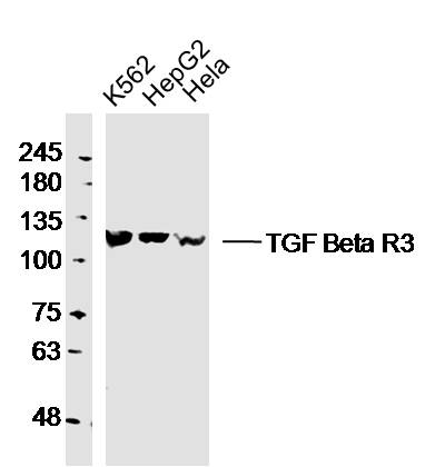
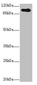
Figure 1. Western blot analysis of TGF Beta Receptor III/TGFBR3 using anti-TGF Beta Receptor III/TGFBR3 antibody (A02765-4). Electrophoresis was performed on a 5-20% SDS-PAGE gel at 70V (Stacking gel) / 90V (Resolving gel) for 2-3 hours. The sample well of each lane was loaded with 30 ug of sample under reducing conditions. Lane 1: human K562 whole cell lysates, Lane 2: human Hela whole cell lysates, Lane 3: rat testis tissue lysates, Lane 4: rat L6 whole cell lysates. After electrophoresis, proteins were transferred to a nitrocellulose membrane at 150 mA for 50-90 minutes. Blocked the membrane with 5% non-fat milk/TBS for 1.5 hour at RT. The membrane was incubated with rabbit anti-TGF Beta Receptor III/TGFBR3 antigen affinity purified polyclonal antibody (Catalog # A02765-4) at 0.25 microg/mL overnight at 4°C, then washed with TBS-0.1%Tween 3 times with 5 minutes each and probed with a goat anti-rabbit IgG-HRP secondary antibody at a dilution of 1:5000 for 1.5 hour at RT. The signal is developed using an Enhanced Chemiluminescent detection (ECL) kit (Catalog # EK1002) with Tanon 5200 system. A specific band was detected for TGF Beta Receptor III/TGFBR3 at approximately 93 kDa. The expected band size for TGF Beta Receptor III/TGFBR3 is at 93 kDa.
Figure 1. Western blot analysis of TGF Beta Receptor III/TGFBR3 using anti-TGF Beta Receptor III/TGFBR3 antibody (A02765-4). Electrophoresis was performed on a 5-20% SDS-PAGE gel at 70V (Stacking gel) / 90V (Resolving gel) for 2-3 hours. The sample well of each lane was loaded with 30 ug of sample under reducing conditions. Lane 1: human K562 whole cell lysates, Lane 2: human Hela whole cell lysates, Lane 3: rat testis tissue lysates, Lane 4: rat L6 whole cell lysates. After electrophoresis, proteins were transferred to a nitrocellulose membrane at 150 mA for 50-90 minutes. Blocked the membrane with 5% non-fat milk/TBS for 1.5 hour at RT. The membrane was incubated with rabbit anti-TGF Beta Receptor III/TGFBR3 antigen affinity purified polyclonal antibody (Catalog # A02765-4) at 0.25 microg/mL overnight at 4°C, then washed with TBS-0.1%Tween 3 times with 5 minutes each and probed with a goat anti-rabbit IgG-HRP secondary antibody at a dilution of 1:5000 for 1.5 hour at RT. The signal is developed using an Enhanced Chemiluminescent detection (ECL) kit (Catalog # EK1002) with Tanon 5200 system. A specific band was detected for TGF Beta Receptor III/TGFBR3 at approximately 93 kDa. The expected band size for TGF Beta Receptor III/TGFBR3 is at 93 kDa.
Anti-TGF beta Receptor III/TGFBR3 Antibody Picoband(r)
A02765-4-FITC
ApplicationsWestern Blot
Product group Antibodies
ReactivityHuman, Rat
TargetTGFBR3
Overview
- SupplierBoster Bio
- Product NameAnti-TGF beta Receptor III/TGFBR3 Antibody Picoband(r)
- Delivery Days Customer9
- ApplicationsWestern Blot
- CertificationResearch Use Only
- ClonalityPolyclonal
- Concentration500 ug/ml
- ConjugateFITC
- Gene ID7049
- Target nameTGFBR3
- Target descriptiontransforming growth factor beta receptor 3
- Target synonymsBGCAN, betaglycan, transforming growth factor beta receptor type 3, TGF-beta receptor type 3, TGF-beta receptor type III, TGFR-3, betaglycan proteoglycan, transforming growth factor beta receptor III
- HostRabbit
- IsotypeIgG
- Protein IDQ03167
- Protein NameTransforming growth factor beta receptor type 3
- Scientific DescriptionBoster Bio Anti-TGF beta Receptor III/TGFBR3 Antibody Picoband® catalog # A02765-4. Tested in WB applications. This antibody reacts with Human, Rat. The brand Picoband indicates this is a premium antibody that guarantees superior quality, high affinity, and strong signals with minimal background in Western blot applications. Only our best-performing antibodies are designated as Picoband, ensuring unmatched performance.
- ReactivityHuman, Rat
- Storage Instruction-20°C,2°C to 8°C
- UNSPSC12352203
Related products
Product group Antibodies
Anti-TGFBR3 Antibody144-61626
ApplicationsImmunoFluorescence, Western Blot
ReactivityHuman, Mouse, Rat
TargetTGFBR3
- SizePrice
Product group Antibodies
ApplicationsImmunoFluorescence, ImmunoCytoChemistry
ReactivityHuman
TargetTGFBR3
- SizePrice
Product group Antibodies

Goat anti-TGFBR3 AntibodyEB08467
ApplicationsFlow Cytometry, ImmunoFluorescence, ELISA
ReactivityHuman
TargetTGFBR3
- SizePrice
Product group Antibodies
Tgfbr3 Polyclonal AntibodyCAC10904
ApplicationsWestern Blot, ELISA, ImmunoHistoChemistry
TargetTGFBR3
- SizePrice
Product group Antibodies
ApplicationsImmunoFluorescence, Western Blot, ELISA, ImmunoCytoChemistry, ImmunoHistoChemistry, ImmunoHistoChemistry Frozen, ImmunoHistoChemistry Paraffin
ReactivityCanine, Chicken, Human, Mouse, Porcine, Rat, Sheep
TargetTGFBR3
- SizePrice
Product group Antibodies
Anti-TGFBR3 AntibodyA121154
ApplicationsFlow Cytometry, ImmunoFluorescence, ELISA
ReactivityHuman
- SizePrice
Product group Antibodies
Anti-TGF beta Receptor III/TGFBR3 Antibody Picoband(r)A02765-4-CARRIER-FREE
ApplicationsWestern Blot
ReactivityHuman, Rat
TargetTGFBR3
- SizePrice
Product group Antibodies
TGFBR3 AntibodyCSB-PA023453ESR1HU
ApplicationsWestern Blot, ELISA, ImmunoHistoChemistry
ReactivityHuman
TargetTGFBR3
- SizePrice