


Immunofluorescent staining of human cell line A549 shows localization to nucleoplasm.

Immunofluorescent staining of human cell line A549 shows localization to nucleoplasm.
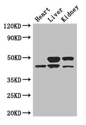
Anti-TGIF1 Antibody
HPA062160
ApplicationsWestern Blot, ChIP Chromatin ImmunoPrecipitation, ImmunoCytoChemistry
Product group Antibodies
ReactivityHuman
TargetTGIF1
Overview
- SupplierAtlas Antibodies
- Product NameAnti-TGIF1 Antibody
- Delivery Days Customer4
- ApplicationsWestern Blot, ChIP Chromatin ImmunoPrecipitation, ImmunoCytoChemistry
- CertificationResearch Use Only
- ClonalityPolyclonal
- ConjugateUnconjugated
- Gene ID7050
- Target nameTGIF1
- Target descriptionTGFB induced factor homeobox 1
- Target synonymsHPE4, TGIF, homeobox protein TGIF1, 5'-TG-3'-interacting factor 1, TALE homeobox TG-interacting factor, transforming growth factor-beta-induced factor
- HostRabbit
- IsotypeIgG
- Protein IDQ15583
- Protein NameHomeobox protein TGIF1
- Scientific DescriptionRecombinant Protein Epitope Signature Tag (PrEST) antigen sequence
- ReactivityHuman
- Storage Instruction-20°C,2°C to 8°C
- UNSPSC41116161
Datasheet
MSDS
Related products
Product group Antibodies
TGIF1 AntibodyCSB-PA623005LA01HU
ApplicationsImmunoFluorescence, Western Blot, ELISA, ImmunoHistoChemistry
ReactivityHuman, Mouse
TargetTGIF1
- SizePrice

Product group Antibodies

Anti-TGIF1 [RAB-T13]Ab01904-1.1
ApplicationsFlow Cytometry, ImmunoFluorescence, ImmunoPrecipitation
ReactivityHuman
TargetTGIF1
- SizePrice

Product group Antibodies
Anti-TGIF/TGIF1 Antibody Picoband(r)A04122-1-CARRIER-FREE
ApplicationsFlow Cytometry, Western Blot, ELISA
ReactivityHuman
TargetTGIF1
- SizePrice
Product group Antibodies

TGIF1 AntibodyLS-C500893
ApplicationsImmunoFluorescence, Western Blot, ELISA, ImmunoHistoChemistry
ReactivityHuman, Mouse
TargetTGIF1
- SizePrice
Product group Antibodies
TGIF1 Polyclonal AntibodyCAC14662
ApplicationsImmunoFluorescence, Western Blot, ELISA, ImmunoHistoChemistry
ReactivityMouse
TargetTGIF1
- SizePrice