


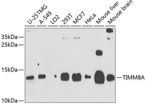
Figure 1. Western blot analysis of TIMM8A/DDP using anti-TIMM8A/DDP antibody (A07659-1). Electrophoresis was performed on a 5-20% SDS-PAGE gel at 70V (Stacking gel) / 90V (Resolving gel) for 2-3 hours. The sample well of each lane was loaded with 50ug of sample under reducing conditions. Lane 1: human HELA whole cell lysates, Lane 2: human MCF-7 whole cell lysates, Lane 3: human PC-3 whole cell lysates, Lane 4: human K562 whole cell lysates, Lane 5: human HEPG2 whole cell lysates, Lane 6: monkey COS-7 whole cell lysates, Lane 7: monkey kidney tissue lysates, Lane 8: rat brain tissue lysates, Lane 9: rat liver tissue lysates, Lane 10: mouse liver tissue lysates. After Electrophoresis, proteins were transferred to a Nitrocellulose membrane at 150mA for 50-90 minutes. Blocked the membrane with 5% Non-fat Milk/ TBS for 1.5 hour at RT. The membrane was incubated with rabbit anti-TIMM8A/DDP antigen affinity purified polyclonal antibody (Catalog # A07659-1) at 0.5 microg/mL overnight at 4°C, then washed with TBS-0.1%Tween 3 times with 5 minutes each and probed with a goat anti-rabbit IgG-HRP secondary antibody at a dilution of 1:5000 for 1.5 hour at RT. The signal is developed using an Enhanced Chemiluminescent detection (ECL) kit (Catalog # EK1002) with Tanon 5200 system. A specific band was detected for TIMM8A/DDP at approximately 13-14KD. The expected band size for TIMM8A/DDP is at 11KD.
. Overlay histogram showing A431 cells stained with A07659-1 (Blue line).The cells were blocked with 10% normal goat serum. And then incubated with rabbit anti-TIMM8A/DDP Antibody (A07659-1, 1microg/1x106 cells) for 30 min at 20°C. DyLight®488 conjugated goat anti-rabbit IgG (BA1127, 5-10microg/1x106 cells) was used as secondary antibody for 30 minutes at 20°C. Isotype control antibody (Green line) was rabbit IgG (1microg/1x106) used under the same conditions. Unlabelled sample (Red line) was also used as a control.")
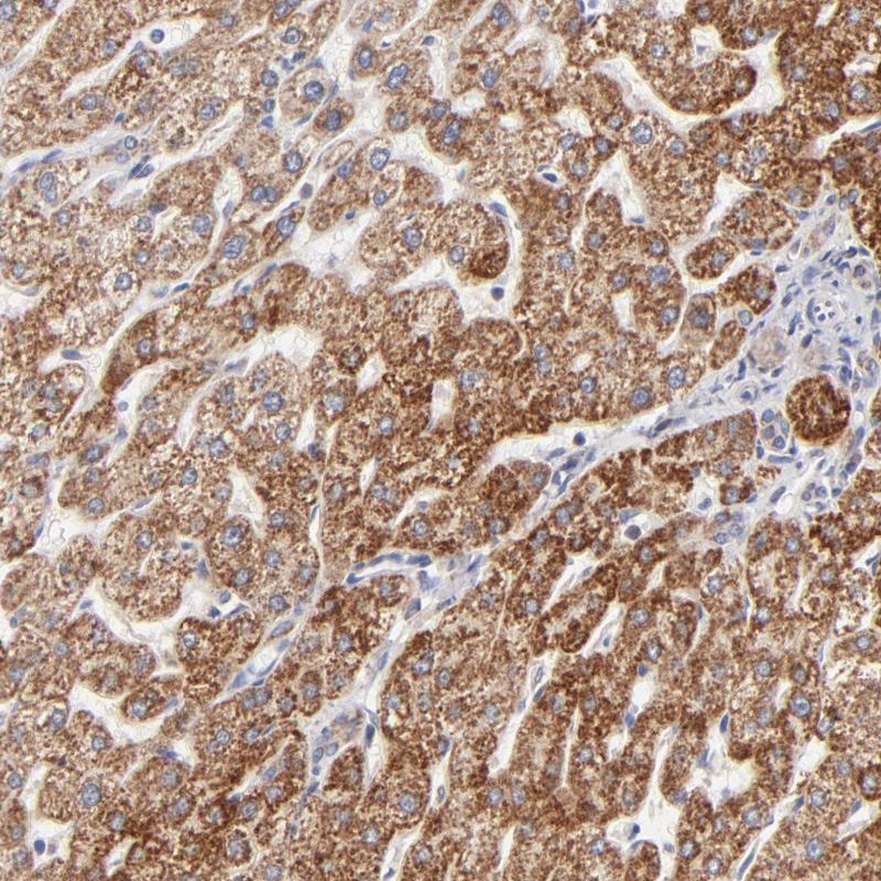
. TIMM8A/DDP was detected in paraffin-embedded section of human melanoma tissue. Heat mediated antigen retrieval was performed in EDTA buffer (pH8.0, epitope retrieval solution). The tissue section was blocked with 10% goat serum. The tissue section was then incubated with 2microg/ml rabbit anti-TIMM8A/DDP Antibody (A07659-1) overnight at 4°C. Biotinylated goat anti-rabbit IgG was used as secondary antibody and incubated for 30 minutes at 37°C. The tissue section was developed using Strepavidin-Biotin-Complex (SABC) (Catalog # SA1022) with DAB as the chromogen.")
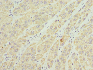
. TIMM8A/DDP was detected in paraffin-embedded section of human testicular cancer tissue. Heat mediated antigen retrieval was performed in EDTA buffer (pH8.0, epitope retrieval solution). The tissue section was blocked with 10% goat serum. The tissue section was then incubated with 2microg/ml rabbit anti-TIMM8A/DDP Antibody (A07659-1) overnight at 4°C. Biotinylated goat anti-rabbit IgG was used as secondary antibody and incubated for 30 minutes at 37°C. The tissue section was developed using Strepavidin-Biotin-Complex (SABC) (Catalog # SA1022) with DAB as the chromogen.")
. TIMM8A/DDP was detected in paraffin-embedded section of human lung cancer tissue. Heat mediated antigen retrieval was performed in EDTA buffer (pH8.0, epitope retrieval solution). The tissue section was blocked with 10% goat serum. The tissue section was then incubated with 2microg/ml rabbit anti-TIMM8A/DDP Antibody (A07659-1) overnight at 4°C. Biotinylated goat anti-rabbit IgG was used as secondary antibody and incubated for 30 minutes at 37°C. The tissue section was developed using Strepavidin-Biotin-Complex (SABC) (Catalog # SA1022) with DAB as the chromogen.")
. TIMM8A/DDP was detected in paraffin-embedded section of human tonsil cancer tissue. Heat mediated antigen retrieval was performed in EDTA buffer (pH8.0, epitope retrieval solution). The tissue section was blocked with 10% goat serum. The tissue section was then incubated with 2microg/ml rabbit anti-TIMM8A/DDP Antibody (A07659-1) overnight at 4°C. Biotinylated goat anti-rabbit IgG was used as secondary antibody and incubated for 30 minutes at 37°C. The tissue section was developed using Strepavidin-Biotin-Complex (SABC) (Catalog # SA1022) with DAB as the chromogen.")
. TIMM8A/DDP was detected in paraffin-embedded section of human bladder cancer tissue. Heat mediated antigen retrieval was performed in EDTA buffer (pH8.0, epitope retrieval solution). The tissue section was blocked with 10% goat serum. The tissue section was then incubated with 2microg/ml rabbit anti-TIMM8A/DDP Antibody (A07659-1) overnight at 4°C. Biotinylated goat anti-rabbit IgG was used as secondary antibody and incubated for 30 minutes at 37°C. The tissue section was developed using Strepavidin-Biotin-Complex (SABC) (Catalog # SA1022) with DAB as the chromogen.")
. TIMM8A/DDP was detected in paraffin-embedded section of human gastric cancer tissue. Heat mediated antigen retrieval was performed in EDTA buffer (pH8.0, epitope retrieval solution). The tissue section was blocked with 10% goat serum. The tissue section was then incubated with 2microg/ml rabbit anti-TIMM8A/DDP Antibody (A07659-1) overnight at 4°C. Biotinylated goat anti-rabbit IgG was used as secondary antibody and incubated for 30 minutes at 37°C. The tissue section was developed using Strepavidin-Biotin-Complex (SABC) (Catalog # SA1022) with DAB as the chromogen.")
. TIMM8A/DDP was detected in paraffin-embedded section of human ovarian cancer tissue. Heat mediated antigen retrieval was performed in EDTA buffer (pH8.0, epitope retrieval solution). The tissue section was blocked with 10% goat serum. The tissue section was then incubated with 2microg/ml rabbit anti-TIMM8A/DDP Antibody (A07659-1) overnight at 4°C. Biotinylated goat anti-rabbit IgG was used as secondary antibody and incubated for 30 minutes at 37°C. The tissue section was developed using Strepavidin-Biotin-Complex (SABC) (Catalog # SA1022) with DAB as the chromogen.")
. TIMM8A/DDP was detected in immunocytochemical section of A431 cells. Enzyme antigen retrieval was performed using IHC enzyme antigen retrieval reagent (AR0022) for 15 mins. The cells were blocked with 10% goat serum. And then incubated with 5microg/mL rabbit anti-TIMM8A/DDP Antibody (A07659-1) overnight at 4°C. DyLight®488 Conjugated Goat Anti-Rabbit IgG (BA1127) was used as secondary antibody at 1:100 dilution and incubated for 30 minutes at 37°C. The section was counterstained with DAPI. Visualize using a fluorescence microscope and filter sets appropriate for the label used.")
Figure 1. Western blot analysis of TIMM8A/DDP using anti-TIMM8A/DDP antibody (A07659-1). Electrophoresis was performed on a 5-20% SDS-PAGE gel at 70V (Stacking gel) / 90V (Resolving gel) for 2-3 hours. The sample well of each lane was loaded with 50ug of sample under reducing conditions. Lane 1: human HELA whole cell lysates, Lane 2: human MCF-7 whole cell lysates, Lane 3: human PC-3 whole cell lysates, Lane 4: human K562 whole cell lysates, Lane 5: human HEPG2 whole cell lysates, Lane 6: monkey COS-7 whole cell lysates, Lane 7: monkey kidney tissue lysates, Lane 8: rat brain tissue lysates, Lane 9: rat liver tissue lysates, Lane 10: mouse liver tissue lysates. After Electrophoresis, proteins were transferred to a Nitrocellulose membrane at 150mA for 50-90 minutes. Blocked the membrane with 5% Non-fat Milk/ TBS for 1.5 hour at RT. The membrane was incubated with rabbit anti-TIMM8A/DDP antigen affinity purified polyclonal antibody (Catalog # A07659-1) at 0.5 microg/mL overnight at 4°C, then washed with TBS-0.1%Tween 3 times with 5 minutes each and probed with a goat anti-rabbit IgG-HRP secondary antibody at a dilution of 1:5000 for 1.5 hour at RT. The signal is developed using an Enhanced Chemiluminescent detection (ECL) kit (Catalog # EK1002) with Tanon 5200 system. A specific band was detected for TIMM8A/DDP at approximately 13-14KD. The expected band size for TIMM8A/DDP is at 11KD.
Anti-TIMM8A/DDP Antibody Picoband(r)
A07659-1-CARRIER-FREE
ApplicationsImmunoFluorescence, Western Blot, ELISA, ImmunoCytoChemistry, ImmunoHistoChemistry
Product group Antibodies
ReactivityHuman, Monkey, Mouse, Rat
TargetTIMM8A
Overview
- SupplierBoster Bio
- Product NameAnti-TIMM8A/DDP Antibody Picoband(r)
- Delivery Days Customer9
- ApplicationsImmunoFluorescence, Western Blot, ELISA, ImmunoCytoChemistry, ImmunoHistoChemistry
- CertificationResearch Use Only
- ClonalityPolyclonal
- Concentration500 ug/ml
- Gene ID1678
- Target nameTIMM8A
- Target descriptiontranslocase of inner mitochondrial membrane 8A
- Target synonymsDDP, DDP1, DFN1, MTS, TIM8, mitochondrial import inner membrane translocase subunit Tim8 A, X-linked deafness dystonia protein, deafness dystonia protein 1, deafness/dystonia peptide, translocase of inner mitochondrial membrane 8 homolog A
- HostRabbit
- IsotypeIgG
- Protein IDO60220
- Protein NameMitochondrial import inner membrane translocase subunit Tim8 A
- Scientific DescriptionBoster Bio Anti-TIMM8A/DDP Antibody Picoband® catalog # A07659-1. Tested in ELISA, IF, IHC, ICC, WB applications. This antibody reacts with Human, Monkey, Mouse, Rat. The brand Picoband indicates this is a premium antibody that guarantees superior quality, high affinity, and strong signals with minimal background in Western blot applications. Only our best-performing antibodies are designated as Picoband, ensuring unmatched performance.
- ReactivityHuman, Monkey, Mouse, Rat
- Storage Instruction-20°C,2°C to 8°C
- UNSPSC12352203
Related products
Product group Antibodies
TIMM8A AntibodyCSB-PA023557ESR2HU
ApplicationsELISA, ImmunoHistoChemistry
ReactivityHuman
TargetTIMM8A
- SizePrice
Product group Antibodies

TIMM8A AntibodyLS-C830542
ApplicationsELISA, ImmunoHistoChemistry
ReactivityHuman, Mouse, Rat
TargetTIMM8A
- SizePrice
Product group Antibodies
Anti-TIMM8A AntibodyHPA003628
ApplicationsImmunoCytoChemistry, ImmunoHistoChemistry
ReactivityHuman
TargetTIMM8A
- SizePrice
Product group Antibodies
TIMM8A antibodyGTX64883
ApplicationsWestern Blot
ReactivityHuman, Mouse
TargetTIMM8A
- SizePrice
Product group Antibodies
Anti-TIMM8A Antibody144-09811
ApplicationsWestern Blot
ReactivityHuman, Mouse
TargetTIMM8A
- SizePrice
Product group Antibodies
TIMM8A Polyclonal AntibodyBS-11769R
ApplicationsImmunoFluorescence, ELISA, ImmunoCytoChemistry, ImmunoHistoChemistry, ImmunoHistoChemistry Frozen, ImmunoHistoChemistry Paraffin
ReactivityBovine, Human, Mouse, Porcine, Rat, Sheep
- SizePrice