


Immunofluorescent staining of human cell line RH-30 shows localization to nucleus.
Immunofluorescent staining of human cell line RH-30 shows localization to nucleus.
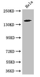
Anti-TNRC6C Antibody
HPA062051
ApplicationsImmunoCytoChemistry
Product group Antibodies
ReactivityHuman
TargetTNRC6C
Overview
- SupplierAtlas Antibodies
- Product NameAnti-TNRC6C Antibody
- Delivery Days Customer4
- ApplicationsImmunoCytoChemistry
- CertificationResearch Use Only
- ClonalityPolyclonal
- ConjugateUnconjugated
- Gene ID57690
- Target nameTNRC6C
- Target descriptiontrinucleotide repeat containing adaptor 6C
- Target synonymstrinucleotide repeat-containing gene 6C protein, trinucleotide repeat containing 6C
- HostRabbit
- IsotypeIgG
- Protein IDQ9HCJ0
- Protein NameTrinucleotide repeat-containing gene 6C protein
- Scientific DescriptionRecombinant Protein Epitope Signature Tag (PrEST) antigen sequence
- ReactivityHuman
- Storage Instruction-20°C,2°C to 8°C
- UNSPSC41116161
Datasheet
MSDS
Related products
Product group Antibodies
TNRC6C Polyclonal AntibodyCAC14622
ApplicationsWestern Blot, ELISA, ImmunoHistoChemistry
TargetTNRC6C
- SizePrice

Product group Antibodies
Anti-TNRC6C Antibody101-11819
ApplicationsImmunoFluorescence, ELISA
TargetTNRC6C
- SizePrice
Product group Antibodies
TNRC6C AntibodyPACO48086
ApplicationsWestern Blot, ELISA, ImmunoHistoChemistry
ReactivityHuman
TargetTNRC6C
- SizePrice
Product group Antibodies

TNRC6C Antibody (Biotin)LS-C501727
ApplicationsELISA
ReactivityHuman
TargetTNRC6C
- SizePrice

Product group Antibodies
Anti-TNRC6C AntibodyHPA022007
ApplicationsImmunoHistoChemistry
ReactivityHuman
TargetTNRC6C
- SizePrice
Product group Antibodies
Anti-TNRC6C AntibodyHPA022007
ApplicationsImmunoHistoChemistry
ReactivityHuman
TargetTNRC6C
- SizePrice
Product group Antibodies
TNRC6C AntibodyCSB-PA862070LA01HU
ApplicationsWestern Blot, ELISA, ImmunoHistoChemistry
ReactivityHuman
TargetTNRC6C
- SizePrice