


Figure 1. Western blot analysis of TOMM20 using anti-TOMM20 antibody (A04039-2). Electrophoresis was performed on a 5-20% SDS-PAGE gel at 70V (Stacking gel) / 90V (Resolving gel) for 2-3 hours. The sample well of each lane was loaded with 30 ug of sample under reducing conditions. Lane 1: human 293T whole cell lysates, Lane 2: human Hela whole cell lysates, Lane 3: human HepG2 whole cell lysates, Lane 4: human Jurkat whole cell lysates, Lane 5: rat brain tissue lysates, Lane 6: rat liver tissue lysates, Lane 7: mouse brain tissue lysates, Lane 8: mouse liver tissue lysates. After electrophoresis, proteins were transferred to a nitrocellulose membrane at 150 mA for 50-90 minutes. Blocked the membrane with 5% non-fat milk/TBS for 1.5 hour at RT. The membrane was incubated with rabbit anti-TOMM20 antigen affinity purified polyclonal antibody (Catalog # A04039-2) at 0.5 microg/mL overnight at 4°C, then washed with TBS-0.1%Tween 3 times with 5 minutes each and probed with a goat anti-rabbit IgG-HRP secondary antibody at a dilution of 1:5000 for 1.5 hour at RT. The signal is developed using an Enhanced Chemiluminescent detection (ECL) kit (Catalog # EK1002) with Tanon 5200 system. A specific band was detected for TOMM20 at approximately 16 kDa. The expected band size for TOMM20 is at 16 kDa.
. TOMM20 was detected in a paraffin-embedded section of human lung cancer tissue. Heat mediated antigen retrieval was performed in EDTA buffer (pH 8.0, epitope retrieval solution). The tissue section was blocked with 10% goat serum. The tissue section was then incubated with 2 microg/ml rabbit anti-TOMM20 Antibody (A04039-2) overnight at 4°C. Peroxidase Conjugated Goat Anti-rabbit IgG was used as secondary antibody and incubated for 30 minutes at 37°C. The tissue section was developed using HRP Conjugated Rabbit IgG Super Vision Assay Kit (Catalog # SV0002) with DAB as the chromogen.")
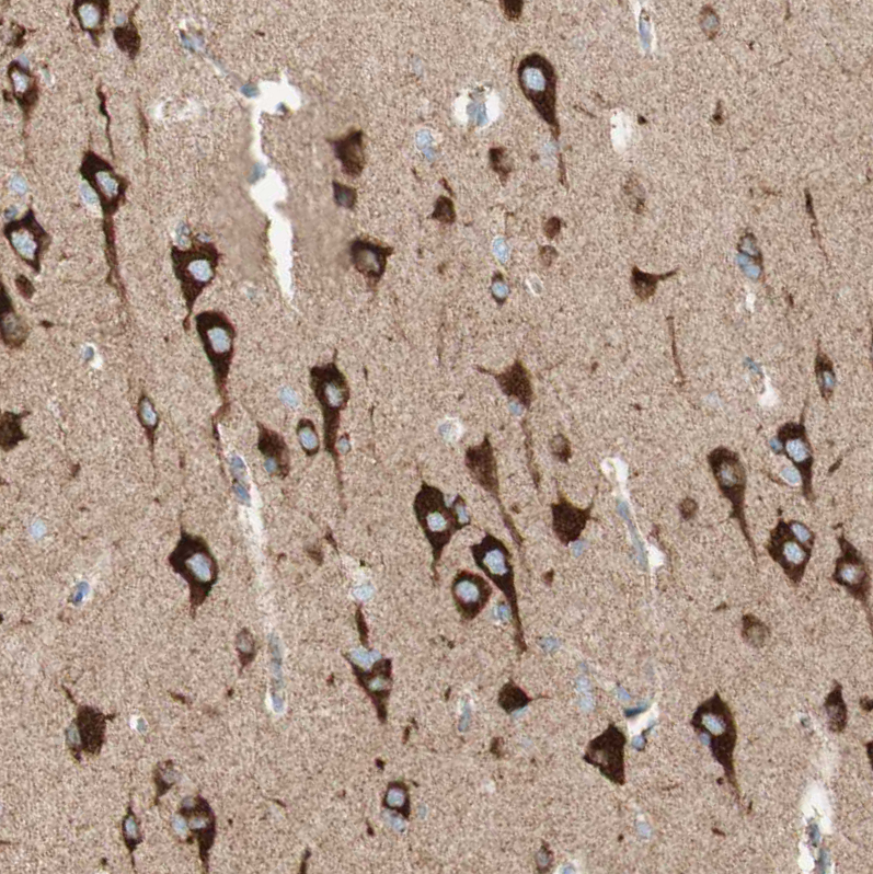
. TOMM20 was detected in a paraffin-embedded section of rat brain tissue. Heat mediated antigen retrieval was performed in EDTA buffer (pH 8.0, epitope retrieval solution). The tissue section was blocked with 10% goat serum. The tissue section was then incubated with 2 microg/ml rabbit anti-TOMM20 Antibody (A04039-2) overnight at 4°C. Peroxidase Conjugated Goat Anti-rabbit IgG was used as secondary antibody and incubated for 30 minutes at 37°C. The tissue section was developed using HRP Conjugated Rabbit IgG Super Vision Assay Kit (Catalog # SV0002) with DAB as the chromogen.")
. TOMM20 was detected in an immunocytochemical section of MG63 cells. Enzyme antigen retrieval was performed using IHC enzyme antigen retrieval reagent (AR0022) for 15 mins. The cells were blocked with 10% goat serum. And then incubated with 5 microg/mL rabbit anti-TOMM20 Antibody (A04039-2) overnight at 4°C. DyLight®488 Conjugated Goat Anti-Rabbit IgG (BA1127) was used as secondary antibody at 1:500 dilution and incubated for 30 minutes at 37°C. The section was counterstained with DAPI. Visualize using a fluorescence microscope and filter sets appropriate for the label used.")
Figure 1. Western blot analysis of TOMM20 using anti-TOMM20 antibody (A04039-2). Electrophoresis was performed on a 5-20% SDS-PAGE gel at 70V (Stacking gel) / 90V (Resolving gel) for 2-3 hours. The sample well of each lane was loaded with 30 ug of sample under reducing conditions. Lane 1: human 293T whole cell lysates, Lane 2: human Hela whole cell lysates, Lane 3: human HepG2 whole cell lysates, Lane 4: human Jurkat whole cell lysates, Lane 5: rat brain tissue lysates, Lane 6: rat liver tissue lysates, Lane 7: mouse brain tissue lysates, Lane 8: mouse liver tissue lysates. After electrophoresis, proteins were transferred to a nitrocellulose membrane at 150 mA for 50-90 minutes. Blocked the membrane with 5% non-fat milk/TBS for 1.5 hour at RT. The membrane was incubated with rabbit anti-TOMM20 antigen affinity purified polyclonal antibody (Catalog # A04039-2) at 0.5 microg/mL overnight at 4°C, then washed with TBS-0.1%Tween 3 times with 5 minutes each and probed with a goat anti-rabbit IgG-HRP secondary antibody at a dilution of 1:5000 for 1.5 hour at RT. The signal is developed using an Enhanced Chemiluminescent detection (ECL) kit (Catalog # EK1002) with Tanon 5200 system. A specific band was detected for TOMM20 at approximately 16 kDa. The expected band size for TOMM20 is at 16 kDa.
Anti-TOMM20 Antibody Picoband(r)
A04039-2-CY3
ApplicationsFlow Cytometry, ImmunoFluorescence, ImmunoPrecipitation, Western Blot, ELISA, ImmunoCytoChemistry, ImmunoHistoChemistry
Product group Antibodies
ReactivityHuman, Mouse, Rat
TargetTOMM20
Overview
- SupplierBoster Bio
- Product NameAnti-TOMM20 Antibody Picoband(r)
- Delivery Days Customer9
- ApplicationsFlow Cytometry, ImmunoFluorescence, ImmunoPrecipitation, Western Blot, ELISA, ImmunoCytoChemistry, ImmunoHistoChemistry
- CertificationResearch Use Only
- ClonalityPolyclonal
- Concentration500 ug/ml
- ConjugateCy3
- Gene ID9804
- Target nameTOMM20
- Target descriptiontranslocase of outer mitochondrial membrane 20
- Target synonymsMAS20, MOM19, TOM20, mitochondrial import receptor subunit TOM20 homolog, mitochondrial 20 kDa outer membrane protein, outer mitochondrial membrane receptor Tom20, translocase of outer mitochondrial membrane 20 homolog type II
- HostRabbit
- IsotypeIgG
- Protein IDQ15388
- Protein NameMitochondrial import receptor subunit TOM20 homolog
- Scientific DescriptionBoster Bio Anti-TOMM20 Antibody Picoband® catalog # A04039-2. Tested in ELISA, Flow Cytometry, IP, IF, IHC, ICC, WB applications. This antibody reacts with Human, Mouse, Rat. The brand Picoband indicates this is a premium antibody that guarantees superior quality, high affinity, and strong signals with minimal background in Western blot applications. Only our best-performing antibodies are designated as Picoband, ensuring unmatched performance.
- ReactivityHuman, Mouse, Rat
- Storage Instruction-20°C,2°C to 8°C
- UNSPSC12352203
Related products
Product group Antibodies
TOMM20 Recombinant AntibodyBSM-61211R
ApplicationsFlow Cytometry, ImmunoFluorescence, Western Blot, ImmunoCytoChemistry, ImmunoHistoChemistry, ImmunoHistoChemistry Frozen, ImmunoHistoChemistry Paraffin
TargetTOMM20
- SizePrice
Product group Antibodies
Tomm20 Polyclonal AntibodyCAC10576
ApplicationsImmunoFluorescence, Western Blot, ELISA, ImmunoHistoChemistry
ReactivityMouse
TargetTOMM20
- SizePrice
Product group Antibodies
Anti-TOMM20 Antibody144-06774
ApplicationsImmunoFluorescence, Western Blot, ImmunoHistoChemistry
ReactivityHuman, Mouse, Rat
TargetTOMM20
- SizePrice

Product group Antibodies
Anti-TOMM20 AntibodyA96710
ApplicationsWestern Blot, ELISA
ReactivityHuman, Mouse, Rat
- SizePrice
Product group Antibodies
References
TOMM20 antibodyGTX133756
ApplicationsImmunoFluorescence, Western Blot, ImmunoCytoChemistry, ImmunoHistoChemistry, ImmunoHistoChemistry Paraffin
ReactivityHuman, Mouse, Rat
TargetTOMM20
- SizePrice
Product group Antibodies

TOMM20 Antibody (30-110 aa, Internal)LS-C386458
ApplicationsWestern Blot, ELISA
ReactivityHuman, Mouse, Rat
TargetTOMM20
- SizePrice
Product group Antibodies
Anti-TOMM20 AntibodyHPA011562
ApplicationsWestern Blot, ImmunoCytoChemistry, ImmunoHistoChemistry
ReactivityHuman
TargetTOMM20
- SizePrice