


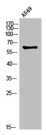
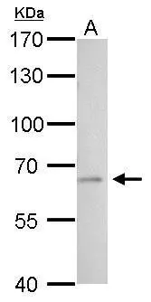
Figure 1. Western blot analysis of PLAT using anti-PLAT antibody (A02965-1). Electrophoresis was performed on a 5-20% SDS-PAGE gel at 70V (Stacking gel) / 90V (Resolving gel) for 2-3 hours. The sample well of each lane was loaded with 50ug of sample under reducing conditions. Lane 1: human Hela whole cell lysates, Lane 2: human U2OS whole cell lysates, Lane 3: human U-87MG whole cell lysates, Lane 4: rat liver tissue lysates, Lane 5: rat kidney tissue lysates, Lane 6: mouse liver tissue lysates, Lane 7: mouse kidney tissue lysates. After Electrophoresis, proteins were transferred to a Nitrocellulose membrane at 150mA for 50-90 minutes. Blocked the membrane with 5% Non-fat Milk/ TBS for 1.5 hour at RT. The membrane was incubated with rabbit anti-PLAT antigen affinity purified polyclonal antibody (Catalog # A02965-1) at 0.5 microg/mL overnight at 4°C, then washed with TBS-0.1%Tween 3 times with 5 minutes each and probed with a goat anti-rabbit IgG-HRP secondary antibody at a dilution of 1:10000 for 1.5 hour at RT. The signal is developed using an Enhanced Chemiluminescent detection (ECL) kit (Catalog # EK1002) with Tanon 5200 system. A specific band was detected for PLAT at approximately 63-69KD. The expected band size for PLAT is at 63KD.
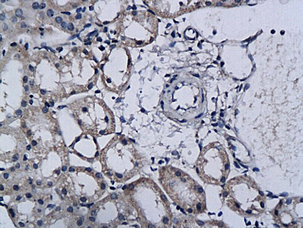
. PLAT was detected in paraffin-embedded section of human placenta tissue. Heat mediated antigen retrieval was performed in citrate buffer (pH6, epitope retrieval solution) for 20 mins. The tissue section was blocked with 10% goat serum. The tissue section was then incubated with 1microg/ml rabbit anti-PLAT Antibody (A02965-1) overnight at 4°C. Biotinylated goat anti-rabbit IgG was used as secondary antibody and incubated for 30 minutes at 37°C. The tissue section was developed using Strepavidin-Biotin-Complex (SABC)(Catalog # SA1022) with DAB as the chromogen.")
. Overlay histogram showing U251 cells stained with A02965-1 (Blue line). To facilitate intracellular staining, cells were fixed with 4% paraformaldehyde and permeabilized with permeabilization buffer. The cells were blocked with 10% normal goat serum. And then incubated with rabbit anti-PLAT Antibody (A02965-1,1microg/1x106 cells) for 30 min at 20°C. DyLight®488 conjugated goat anti-rabbit IgG (BA1127, 5-10microg/1x106 cells) was used as secondary antibody for 30 minutes at 20°C. Isotype control antibody (Green line) was rabbit IgG (1microg/1x106) used under the same conditions. Unlabelled sample (Red line) was also used as a control.")
Figure 1. Western blot analysis of PLAT using anti-PLAT antibody (A02965-1). Electrophoresis was performed on a 5-20% SDS-PAGE gel at 70V (Stacking gel) / 90V (Resolving gel) for 2-3 hours. The sample well of each lane was loaded with 50ug of sample under reducing conditions. Lane 1: human Hela whole cell lysates, Lane 2: human U2OS whole cell lysates, Lane 3: human U-87MG whole cell lysates, Lane 4: rat liver tissue lysates, Lane 5: rat kidney tissue lysates, Lane 6: mouse liver tissue lysates, Lane 7: mouse kidney tissue lysates. After Electrophoresis, proteins were transferred to a Nitrocellulose membrane at 150mA for 50-90 minutes. Blocked the membrane with 5% Non-fat Milk/ TBS for 1.5 hour at RT. The membrane was incubated with rabbit anti-PLAT antigen affinity purified polyclonal antibody (Catalog # A02965-1) at 0.5 microg/mL overnight at 4°C, then washed with TBS-0.1%Tween 3 times with 5 minutes each and probed with a goat anti-rabbit IgG-HRP secondary antibody at a dilution of 1:10000 for 1.5 hour at RT. The signal is developed using an Enhanced Chemiluminescent detection (ECL) kit (Catalog # EK1002) with Tanon 5200 system. A specific band was detected for PLAT at approximately 63-69KD. The expected band size for PLAT is at 63KD.
Anti-TPA Tissue Plasminogen Activator/PLAT Antibody Picoband(r)
A02965-1-CARRIER-FREE
ApplicationsFlow Cytometry, Western Blot, ImmunoCytoChemistry, ImmunoHistoChemistry, ImmunoHistoChemistry Frozen
Product group Antibodies
ReactivityHuman, Mouse, Rat
TargetPLAT
Overview
- SupplierBoster Bio
- Product NameAnti-TPA Tissue Plasminogen Activator/PLAT Antibody Picoband(r)
- Delivery Days Customer9
- ApplicationsFlow Cytometry, Western Blot, ImmunoCytoChemistry, ImmunoHistoChemistry, ImmunoHistoChemistry Frozen
- CertificationResearch Use Only
- ClonalityPolyclonal
- Concentration500 ug/ml
- Gene ID5327
- Target namePLAT
- Target descriptionplasminogen activator, tissue type
- Target synonymsT-PA, TPA, tissue-type plasminogen activator, alteplase, plasminogen/activator kringle, reteplase, t-plasminogen activator
- HostRabbit
- IsotypeIgG
- Protein IDP00750
- Protein NameTissue-type plasminogen activator
- Scientific DescriptionBoster Bio Anti-TPA Tissue Plasminogen Activator/PLAT Antibody Picoband® catalog # A02965-1. Tested in Flow Cytometry, IHC, IHC-F, ICC, WB applications. This antibody reacts with Human, Mouse, Rat. The brand Picoband indicates this is a premium antibody that guarantees superior quality, high affinity, and strong signals with minimal background in Western blot applications. Only our best-performing antibodies are designated as Picoband, ensuring unmatched performance.
- ReactivityHuman, Mouse, Rat
- Storage Instruction-20°C,2°C to 8°C
- UNSPSC12352203
Related products
Product group Antibodies
Anti-tPA AntibodyA98425
ApplicationsWestern Blot, ELISA
ReactivityHuman, Mouse, Rat
- SizePrice
Product group Antibodies
TPA Polyclonal AntibodyBS-1545R
ApplicationsImmunoFluorescence, Western Blot, ELISA, ImmunoCytoChemistry, ImmunoHistoChemistry, ImmunoHistoChemistry Frozen, ImmunoHistoChemistry Paraffin
ReactivityBovine, Equine, Human, Mouse, Porcine, Rabbit, Rat
TargetPLAT
- SizePrice
Product group Antibodies
Plat Polyclonal AntibodyCAC09151
ApplicationsWestern Blot, ELISA, ImmunoHistoChemistry
ReactivityMouse
TargetPLAT
- SizePrice
Product group Antibodies
PLAT AntibodyCSB-PA030068
ApplicationsWestern Blot, ELISA
ReactivityHuman, Mouse, Rat
TargetPLAT
- SizePrice
Product group Antibodies

PLAT / TPA AntibodyLS-C402793
ApplicationsWestern Blot, ELISA, ImmunoHistoChemistry
ReactivityHuman
TargetPLAT
- SizePrice
Product group Antibodies
tPA antibodyGTX103453
ApplicationsWestern Blot, ImmunoHistoChemistry, ImmunoHistoChemistry Paraffin
ReactivityHuman, Mouse, Rat
TargetPLAT
- SizePrice

Product group Antibodies
Anti-PLAT AntibodyHPA003412
ApplicationsImmunoHistoChemistry
ReactivityHuman
TargetPLAT
- SizePrice
Product group Antibodies
TargetPLAT
- SizePrice
Product group Antibodies
Anti-PLATY158213
ApplicationsWestern Blot, ELISA, ImmunoHistoChemistry
ReactivityHuman
- SizePrice