


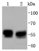
Figure 1. Western blot analysis of TRK fused gene/TFG using anti-TRK fused gene/TFG antibody (A02870-3). Electrophoresis was performed on a 5-20% SDS-PAGE gel at 70V (Stacking gel) / 90V (Resolving gel) for 2-3 hours. The sample well of each lane was loaded with 30 ug of sample under reducing conditions. Lane 1: human MCF-7 whole cell lysates, Lane 2: human Hela whole cell lysates, Lane 3: human A549 whole cell lysates, Lane 4: human PC-3 whole cell lysates, Lane 5: human A431 whole cell lysates, Lane 6: human U-87MG whole cell lysates, Lane 7: human Hacat whole cell lysates, Lane 8: human T-47D whole cell lysates, Lane 9: rat pancreas tissue lysates, Lane 10: rat PC-12 whole cell lysates, Lane 11: mouse small intestine tissue lysates, Lane 12: mouse HEPA1-6 whole cell lysates. After electrophoresis, proteins were transferred to a nitrocellulose membrane at 150 mA for 50-90 minutes. Blocked the membrane with 5% non-fat milk/TBS for 1.5 hour at RT. The membrane was incubated with rabbit anti-TRK fused gene/TFG antigen affinity purified polyclonal antibody (Catalog # A02870-3) at 0.25 microg/mL overnight at 4°C, then washed with TBS-0.1%Tween 3 times with 5 minutes each and probed with a goat anti-rabbit IgG-HRP secondary antibody at a dilution of 1:5000 for 1.5 hour at RT. The signal is developed using an Enhanced Chemiluminescent detection (ECL) kit (Catalog # EK1002) with Tanon 5200 system. A specific band was detected for TRK fused gene/TFG at approximately 60 kDa. The expected band size for TRK fused gene/TFG is at 43 kDa.
. TRK fused gene/TFG was detected in a paraffin-embedded section of human breast cancer tissue. Heat mediated antigen retrieval was performed in EDTA buffer (pH 8.0, epitope retrieval solution). The tissue section was blocked with 10% goat serum. The tissue section was then incubated with 2 microg/ml rabbit anti-TRK fused gene/TFG Antibody (A02870-3) overnight at 4°C. Peroxidase Conjugated Goat Anti-rabbit IgG was used as secondary antibody and incubated for 30 minutes at 37°C. The tissue section was developed using HRP Conjugated Rabbit IgG Super Vision Assay Kit (Catalog # SV0002) with DAB as the chromogen.")
. TRK fused gene/TFG was detected in a paraffin-embedded section of human cervical cancer tissue. Heat mediated antigen retrieval was performed in EDTA buffer (pH 8.0, epitope retrieval solution). The tissue section was blocked with 10% goat serum. The tissue section was then incubated with 2 microg/ml rabbit anti-TRK fused gene/TFG Antibody (A02870-3) overnight at 4°C. Peroxidase Conjugated Goat Anti-rabbit IgG was used as secondary antibody and incubated for 30 minutes at 37°C. The tissue section was developed using HRP Conjugated Rabbit IgG Super Vision Assay Kit (Catalog # SV0002) with DAB as the chromogen.")
. TRK fused gene/TFG was detected in a paraffin-embedded section of human liver cancer tissue. Heat mediated antigen retrieval was performed in EDTA buffer (pH 8.0, epitope retrieval solution). The tissue section was blocked with 10% goat serum. The tissue section was then incubated with 2 microg/ml rabbit anti-TRK fused gene/TFG Antibody (A02870-3) overnight at 4°C. Peroxidase Conjugated Goat Anti-rabbit IgG was used as secondary antibody and incubated for 30 minutes at 37°C. The tissue section was developed using HRP Conjugated Rabbit IgG Super Vision Assay Kit (Catalog # SV0002) with DAB as the chromogen.")
. TRK fused gene/TFG was detected in a paraffin-embedded section of human lung tissue. Heat mediated antigen retrieval was performed in EDTA buffer (pH 8.0, epitope retrieval solution). The tissue section was blocked with 10% goat serum. The tissue section was then incubated with 2 microg/ml rabbit anti-TRK fused gene/TFG Antibody (A02870-3) overnight at 4°C. Peroxidase Conjugated Goat Anti-rabbit IgG was used as secondary antibody and incubated for 30 minutes at 37°C. The tissue section was developed using HRP Conjugated Rabbit IgG Super Vision Assay Kit (Catalog # SV0002) with DAB as the chromogen.")
. TRK fused gene/TFG was detected in a paraffin-embedded section of human ovarian cancer tissue. Heat mediated antigen retrieval was performed in EDTA buffer (pH 8.0, epitope retrieval solution). The tissue section was blocked with 10% goat serum. The tissue section was then incubated with 2 microg/ml rabbit anti-TRK fused gene/TFG Antibody (A02870-3) overnight at 4°C. Peroxidase Conjugated Goat Anti-rabbit IgG was used as secondary antibody and incubated for 30 minutes at 37°C. The tissue section was developed using HRP Conjugated Rabbit IgG Super Vision Assay Kit (Catalog # SV0002) with DAB as the chromogen.")
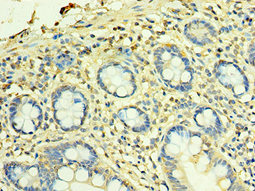
. TRK fused gene/TFG was detected in a paraffin-embedded section of rat colon tissue. Heat mediated antigen retrieval was performed in EDTA buffer (pH 8.0, epitope retrieval solution). The tissue section was blocked with 10% goat serum. The tissue section was then incubated with 2 microg/ml rabbit anti-TRK fused gene/TFG Antibody (A02870-3) overnight at 4°C. Peroxidase Conjugated Goat Anti-rabbit IgG was used as secondary antibody and incubated for 30 minutes at 37°C. The tissue section was developed using HRP Conjugated Rabbit IgG Super Vision Assay Kit (Catalog # SV0002) with DAB as the chromogen.")
. TRK fused gene/TFG was detected in an immunocytochemical section of U87 cells. Enzyme antigen retrieval was performed using IHC enzyme antigen retrieval reagent (AR0022) for 15 mins. The cells were blocked with 10% goat serum. And then incubated with 5 microg/mL rabbit anti-TRK fused gene/TFG Antibody (A02870-3) overnight at 4°C. DyLight®488 Conjugated Goat Anti-Rabbit IgG (BA1127) was used as secondary antibody at 1:100 dilution and incubated for 30 minutes at 37°C. The section was counterstained with DAPI. Visualize using a fluorescence microscope and filter sets appropriate for the label used.")
. TRK fused gene/TFG was detected in a paraffin-embedded section of human colon cancer tissue. Heat mediated antigen retrieval was performed in EDTA buffer (pH 8.0, epitope retrieval solution). The tissue section was blocked with 10% goat serum. The tissue section was then incubated with 5 microg/mL rabbit anti-TRK fused gene/TFG Antibody (A02870-3) overnight at 4°C. DyLight®550 Conjugated Goat Anti-Rabbit IgG (BA1135) was used as secondary antibody at 1:500 dilution and incubated for 30 minutes at 37°C. The section was counterstained with DAPI. Visualize using a fluorescence microscope and filter sets appropriate for the label used.")
Figure 1. Western blot analysis of TRK fused gene/TFG using anti-TRK fused gene/TFG antibody (A02870-3). Electrophoresis was performed on a 5-20% SDS-PAGE gel at 70V (Stacking gel) / 90V (Resolving gel) for 2-3 hours. The sample well of each lane was loaded with 30 ug of sample under reducing conditions. Lane 1: human MCF-7 whole cell lysates, Lane 2: human Hela whole cell lysates, Lane 3: human A549 whole cell lysates, Lane 4: human PC-3 whole cell lysates, Lane 5: human A431 whole cell lysates, Lane 6: human U-87MG whole cell lysates, Lane 7: human Hacat whole cell lysates, Lane 8: human T-47D whole cell lysates, Lane 9: rat pancreas tissue lysates, Lane 10: rat PC-12 whole cell lysates, Lane 11: mouse small intestine tissue lysates, Lane 12: mouse HEPA1-6 whole cell lysates. After electrophoresis, proteins were transferred to a nitrocellulose membrane at 150 mA for 50-90 minutes. Blocked the membrane with 5% non-fat milk/TBS for 1.5 hour at RT. The membrane was incubated with rabbit anti-TRK fused gene/TFG antigen affinity purified polyclonal antibody (Catalog # A02870-3) at 0.25 microg/mL overnight at 4°C, then washed with TBS-0.1%Tween 3 times with 5 minutes each and probed with a goat anti-rabbit IgG-HRP secondary antibody at a dilution of 1:5000 for 1.5 hour at RT. The signal is developed using an Enhanced Chemiluminescent detection (ECL) kit (Catalog # EK1002) with Tanon 5200 system. A specific band was detected for TRK fused gene/TFG at approximately 60 kDa. The expected band size for TRK fused gene/TFG is at 43 kDa.
Anti-TRK fused gene/TFG Antibody Picoband(r)
A02870-3-DYLIGHT488
ApplicationsImmunoFluorescence, Western Blot, ELISA, ImmunoCytoChemistry, ImmunoHistoChemistry
Product group Antibodies
ReactivityHuman, Mouse, Rat
TargetTFG
Overview
- SupplierBoster Bio
- Product NameAnti-TRK fused gene/TFG Antibody Picoband(r)
- Delivery Days Customer9
- ApplicationsImmunoFluorescence, Western Blot, ELISA, ImmunoCytoChemistry, ImmunoHistoChemistry
- CertificationResearch Use Only
- ClonalityPolyclonal
- Concentration500 ug/ml
- ConjugateDyLight 488
- Gene ID10342
- Target nameTFG
- Target descriptiontrafficking from ER to golgi regulator
- Target synonymsHMSNP, SPG57, TF6, TRKT3, protein TFG, TRK-fused, TRK-fused gene protein, TRKT3 oncogene
- HostRabbit
- IsotypeIgG
- Protein IDQ92734
- Protein NameProtein TFG
- Scientific DescriptionBoster Bio Anti-TRK fused gene/TFG Antibody Picoband® catalog # A02870-3. Tested in ELISA, IF, IHC, ICC, WB applications. This antibody reacts with Human, Mouse, Rat. The brand Picoband indicates this is a premium antibody that guarantees superior quality, high affinity, and strong signals with minimal background in Western blot applications. Only our best-performing antibodies are designated as Picoband, ensuring unmatched performance.
- ReactivityHuman, Mouse, Rat
- Storage Instruction-20°C,2°C to 8°C
- UNSPSC12352203
Related products
Product group Antibodies
Anti-TFG Antibody144-07769
ApplicationsWestern Blot, ImmunoHistoChemistry
ReactivityHuman, Mouse, Rat
TargetTFG
- SizePrice
Product group Antibodies
Anti-TRK fused gene/TFG Antibody Picoband(r)A02870-3-CARRIER-FREE
ApplicationsImmunoFluorescence, Western Blot, ELISA, ImmunoCytoChemistry, ImmunoHistoChemistry
ReactivityHuman, Mouse, Rat
TargetTFG
- SizePrice
Product group Antibodies
TFG Recombinant AntibodyBSM-52676R
ApplicationsImmunoFluorescence, Western Blot, ImmunoCytoChemistry, ImmunoHistoChemistry, ImmunoHistoChemistry Frozen, ImmunoHistoChemistry Paraffin
ReactivityHuman, Mouse, Rat
TargetTFG
- SizePrice
![WB analysis of Jurkat cell lysate using GTX00523 TFG antibody [TFG-03].](https://www.genetex.com/upload/website/prouct_img/normal/GTX00523/GTX00523_20191025_AP_001_61_w_23053121_839.webp)
Product group Antibodies
TFG antibody [TFG-03]GTX00523
ApplicationsImmunoFluorescence, ImmunoPrecipitation, Western Blot, ImmunoCytoChemistry
ReactivityHuman
TargetTFG
- SizePrice
Product group Antibodies

TFG AntibodyLS-C409319
ApplicationsWestern Blot, ImmunoHistoChemistry
ReactivityHuman, Mouse, Rat
TargetTFG
- SizePrice
Product group Antibodies
TFG AntibodyCSB-PA852902ESR1HU
ApplicationsELISA, ImmunoHistoChemistry
ReactivityHuman
TargetTFG
- SizePrice

Product group Antibodies
Anti-TFG AntibodyHPA052206
ApplicationsWestern Blot, ImmunoCytoChemistry
ReactivityHuman
TargetTFG
- SizePrice