


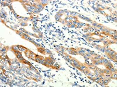
Anti-TRPA1 Antibody
A46164
ApplicationsImmunoHistoChemistry
Product group Antibodies
ReactivityHuman
Overview
- SupplierAntibodies.com
- Product NameAnti-TRPA1 Antibody
- Delivery Days Customer7
- ApplicationsImmunoHistoChemistry
- CertificationResearch Use Only
- ClonalityPolyclonal
- Concentration2 mg/ml
- ConjugateUnconjugated
- HostRabbit
- Scientific DescriptionRabbit polyclonal antibody to TRPA1
- ReactivityHuman
- UNSPSC12352203
Related products
Product group Antibodies
Anti-TRPA1 Antibody144-08568
ApplicationsImmunoFluorescence, Western Blot, ImmunoHistoChemistry
ReactivityHuman, Mouse, Rat
TargetTRPA1
- SizePrice

Product group Antibodies
Anti-TRPA1/TSA Antibody Picoband(r)A00453-2-CARRIER-FREE
ApplicationsFlow Cytometry, Western Blot, ELISA
ReactivityHuman
TargetTRPA1
- SizePrice
Product group Antibodies
TRPA1 AntibodyCSB-PA912872
ApplicationsELISA, ImmunoHistoChemistry
ReactivityHuman
TargetTRPA1
- SizePrice
Product group Antibodies
Transient Receptor Potential Cation Channel Subfamily A, Member 1 (TRPA1) Polyclonal AntibodyCAU22389
ApplicationsWestern Blot, ELISA, ImmunoCytoChemistry, ImmunoHistoChemistry, ImmunoHistoChemistry Frozen, ImmunoHistoChemistry Paraffin
ReactivityPorcine
TargetTRPA1
- SizePrice
Product group Antibodies

TRPA1 AntibodyLS-C406070
ApplicationsELISA, ImmunoHistoChemistry
ReactivityHuman
TargetTRPA1
- SizePrice

Product group Antibodies
References
TRPA1 antibodyGTX54765
ApplicationsImmunoFluorescence, ImmunoPrecipitation, Western Blot, ImmunoCytoChemistry, ImmunoHistoChemistry, ImmunoHistoChemistry Frozen, ImmunoHistoChemistry Paraffin, Other Application
ReactivityHuman, Mouse, Rat
TargetTRPA1
- SizePrice
Product group Antibodies
Anti-TRPA1 AntibodyCAB8568
ApplicationsImmunoFluorescence, Western Blot, ELISA, ImmunoCytoChemistry, ImmunoHistoChemistry, ImmunoHistoChemistry Paraffin
ReactivityHuman
TargetTRPA1
- SizePrice