


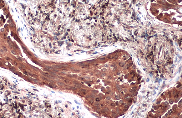
Immunohistochemical staining of human fallopian tube shows strong cytoplasmic positivity in glandular cells.

Immunohistochemical staining of human fallopian tube shows strong cytoplasmic positivity in glandular cells.
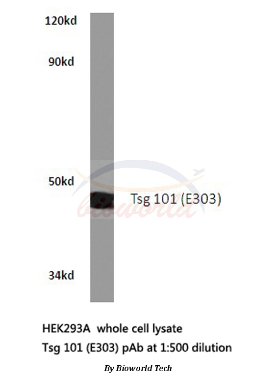
Anti-TSG101 Antibody
HPA006161
ApplicationsWestern Blot, ImmunoCytoChemistry, ImmunoHistoChemistry
Product group Antibodies
ReactivityHuman, Mouse, Rat
TargetTSG101
Overview
- SupplierAtlas Antibodies
- Product NameAnti-TSG101 Antibody
- Delivery Days Customer4
- ApplicationsWestern Blot, ImmunoCytoChemistry, ImmunoHistoChemistry
- CertificationResearch Use Only
- ClonalityPolyclonal
- ConjugateUnconjugated
- Gene ID7251
- Target nameTSG101
- Target descriptiontumor susceptibility 101
- Target synonymsTSG10, VPS23, tumor susceptibility gene 101 protein, ESCRT-I complex subunit TSG101, tumor susceptibility gene 10, tumor susceptibility gene 101, tumor susceptibility protein
- HostRabbit
- IsotypeIgG
- Protein IDQ99816
- Protein NameTumor susceptibility gene 101 protein
- Scientific DescriptionRecombinant Protein Epitope Signature Tag (PrEST) antigen sequence
- ReactivityHuman, Mouse, Rat
- Storage Instruction-20°C,2°C to 8°C
- UNSPSC41116161
Datasheet
MSDS
Related products
Product group Antibodies
ApplicationsImmunoFluorescence, Western Blot, ImmunoHistoChemistry
ReactivityHuman, Mouse, Rat
- SizePrice

Product group Antibodies
Anti-TSG101 Antibody Picoband(r)A01233-2-CARRIER-FREE
ApplicationsFlow Cytometry, Western Blot, ELISA
ReactivityHuman, Mouse, Rat
TargetTSG101
- SizePrice
Product group Antibodies
Anti-TSG101 Antibody144-61333
ApplicationsImmunoPrecipitation, Western Blot
ReactivityHuman, Mouse, Rat
TargetTSG101
- SizePrice
Product group Antibodies

TSG101 AntibodyLS-C761188
ApplicationsImmunoFluorescence, Western Blot, ImmunoCytoChemistry, ImmunoHistoChemistry
ReactivityHuman, Mouse, Rat
TargetTSG101
- SizePrice

Product group Antibodies
References
Tsg101 Polyclonal AntibodyBS-1365R
ApplicationsImmunoFluorescence, Western Blot, ELISA, ImmunoCytoChemistry, ImmunoHistoChemistry, ImmunoHistoChemistry Frozen, ImmunoHistoChemistry Paraffin
ReactivityBovine, Canine, Equine, Human, Mouse, Rabbit, Rat
TargetTSG101
- SizePrice
Product group Antibodies
TSG101 AntibodyCSB-PA060017
ApplicationsImmunoFluorescence, Western Blot, ELISA, ImmunoHistoChemistry
ReactivityHuman, Monkey, Mouse, Rat
TargetTSG101
- SizePrice
Product group Antibodies
ApplicationsImmunoPrecipitation, Western Blot, ImmunoCytoChemistry, ImmunoHistoChemistry
ReactivityMouse, Porcine
TargetTSG101
- SizePrice
Product group Antibodies
TSG101 antibodyGTX118736
ApplicationsImmunoPrecipitation, Western Blot, ELISA, ImmunoHistoChemistry, ImmunoHistoChemistry Paraffin
ReactivityHuman, Mouse, Rat
TargetTSG101
- SizePrice