


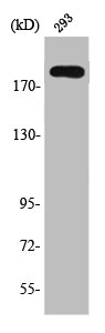
Figure 1. Western blot analysis of TSC2 using anti-TSC2 antibody (M00229-1). Electrophoresis was performed on a 5-20% SDS-PAGE gel at 70V (Stacking gel) / 90V (Resolving gel) for 2-3 hours. The sample well of each lane was loaded with 50ug of sample under reducing conditions. Lane 1: human HEK293 tissue lysates, Lane 2: human PC-3 whole cell lysates, After Electrophoresis, proteins were transferred to a Nitrocellulose membrane at 150mA for 50-90 minutes. Blocked the membrane with 5% Non-fat Milk/ TBS for 1.5 hour at RT. The membrane was incubated with mouse anti-TSC2 antigen affinity purified polyclonal antibody (Catalog # M00229-1) at 0.5 microg/mL overnight at 4°C, then washed with TBS-0.1%Tween 3 times with 5 minutes each and probed with a goat anti-mouse IgG-HRP secondary antibody at a dilution of 1:10000 for 1.5 hour at RT. The signal is developed using an Enhanced Chemiluminescent detection (ECL) kit (Catalog # EK1001) with Tanon 5200 system. A specific band was detected for TSC2 at approximately 280KD. The expected band size for TSC2 is at 201KD.
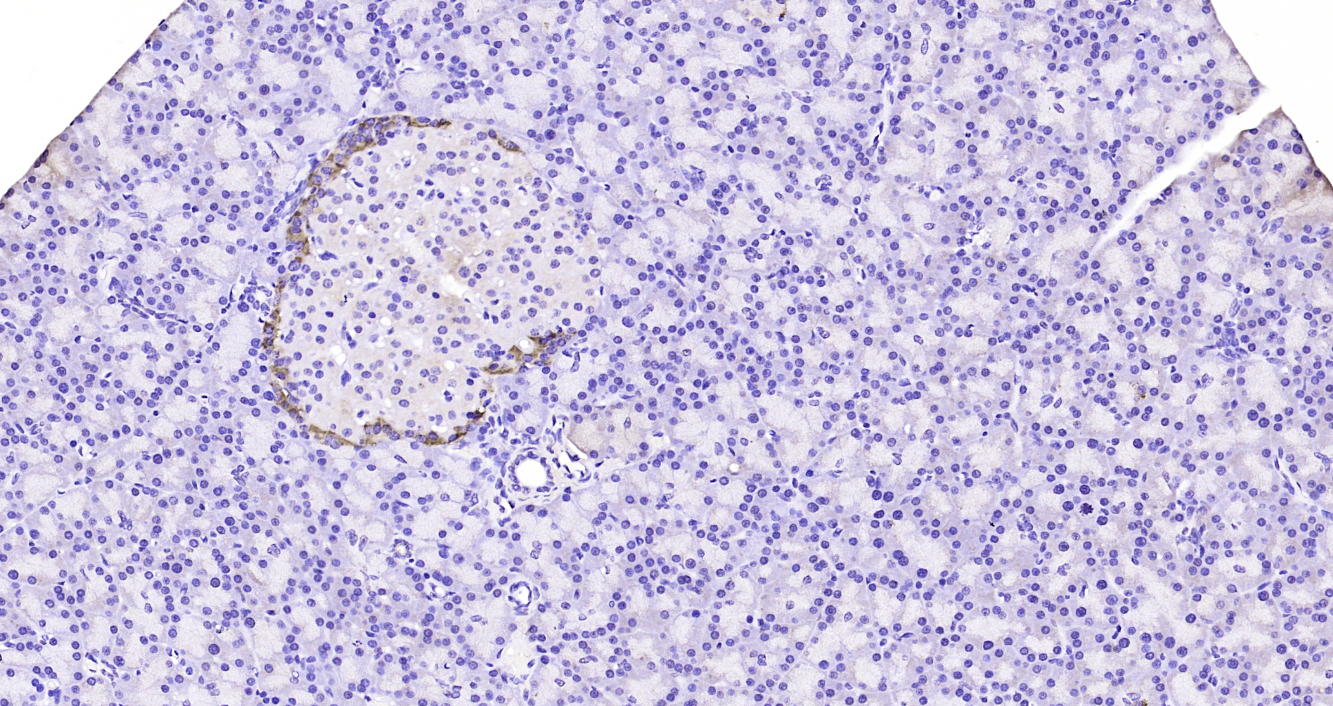
. TSC2 was detected in a paraffin-embedded section of human liver cancer tissue. Heat mediated antigen retrieval was performed in EDTA buffer (pH 8.0, epitope retrieval solution). The tissue section was blocked with 10% goat serum. The tissue section was then incubated with 1 microg/ml mouse anti-TSC2 Antibody (M00229-1) overnight at 4°C. Peroxidase Conjugated Goat Anti-mouse IgG was used as secondary antibody and incubated for 30 minutes at 37°C. The tissue section was developed using HRP Conjugated Mouse IgG Super Vision Assay Kit (Catalog # SV0001) with DAB as the chromogen.")
. Overlay histogram showing A549 cells stained with M00229-1 (Blue line). To facilitate intracellular staining, cells were fixed with 4% paraformaldehyde and permeabilized with permeabilization buffer. The cells were blocked with 10% normal goat serum. And then incubated with mouse anti-TSC2 Antibody (M00229-1, 1 microg/1x106 cells) for 30 min at 20°C. DyLight®488 conjugated goat anti-mouse IgG (BA1126, 5-10 microg/1x106 cells) was used as secondary antibody for 30 minutes at 20°C. Isotype control antibody (Green line) was mouse IgG (1 microg/1x106) used under the same conditions. Unlabelled sample (Red line) was also used as a control.")
Figure 1. Western blot analysis of TSC2 using anti-TSC2 antibody (M00229-1). Electrophoresis was performed on a 5-20% SDS-PAGE gel at 70V (Stacking gel) / 90V (Resolving gel) for 2-3 hours. The sample well of each lane was loaded with 50ug of sample under reducing conditions. Lane 1: human HEK293 tissue lysates, Lane 2: human PC-3 whole cell lysates, After Electrophoresis, proteins were transferred to a Nitrocellulose membrane at 150mA for 50-90 minutes. Blocked the membrane with 5% Non-fat Milk/ TBS for 1.5 hour at RT. The membrane was incubated with mouse anti-TSC2 antigen affinity purified polyclonal antibody (Catalog # M00229-1) at 0.5 microg/mL overnight at 4°C, then washed with TBS-0.1%Tween 3 times with 5 minutes each and probed with a goat anti-mouse IgG-HRP secondary antibody at a dilution of 1:10000 for 1.5 hour at RT. The signal is developed using an Enhanced Chemiluminescent detection (ECL) kit (Catalog # EK1001) with Tanon 5200 system. A specific band was detected for TSC2 at approximately 280KD. The expected band size for TSC2 is at 201KD.
Anti-Tuberin TSC2 Antibody Picoband(r) (monoclonal, 6I3)
M00229-1-DYLIGHT488
ApplicationsFlow Cytometry, Western Blot, ImmunoHistoChemistry
Product group Antibodies
ReactivityHuman
TargetTSC2
Overview
- SupplierBoster Bio
- Product NameAnti-Tuberin TSC2 Antibody Picoband(r) (monoclonal, 6I3)
- Delivery Days Customer9
- ApplicationsFlow Cytometry, Western Blot, ImmunoHistoChemistry
- CertificationResearch Use Only
- ClonalityMonoclonal
- Clone ID6I3
- ConjugateDyLight 488
- Gene ID7249
- Target nameTSC2
- Target descriptionTSC complex subunit 2
- Target synonymsLAM, PPP1R160, TSC4, tuberin, protein phosphatase 1, regulatory subunit 160, tuberous sclerosis 2 protein
- HostMouse
- IsotypeIgG2b
- Protein IDP49815
- Protein NameTuberin
- Scientific DescriptionBoster Bio Anti-Tuberin TSC2 Antibody Picoband® (monoclonal, 6I3) catalog # M00229-1. Tested in Flow Cytometry, IHC, WB applications. This antibody reacts with Human. The brand Picoband indicates this is a premium antibody that guarantees superior quality, high affinity, and strong signals with minimal background in Western blot applications. Only our best-performing antibodies are designated as Picoband, ensuring unmatched performance.
- ReactivityHuman
- Storage Instruction-20°C,2°C to 8°C
- UNSPSC12352203
Related products
Product group Antibodies

Anti-TSC2 [RAB-C431]Ab01910-1.1
ApplicationsImmunoPrecipitation
ReactivityHuman
TargetTSC2
- SizePrice
Product group Antibodies
Anti-TSC2 Antibody144-61623
ApplicationsWestern Blot, ImmunoHistoChemistry
ReactivityHuman, Mouse, Rat
TargetTSC2
- SizePrice
Product group Antibodies
TSC2 Polyclonal AntibodyCAC14612
ApplicationsImmunoFluorescence, Western Blot, ELISA
ReactivityMouse
TargetTSC2
- SizePrice
Product group Antibodies
References
Tuberin Polyclonal AntibodyBS-3586R
ApplicationsImmunoFluorescence, ELISA, ImmunoCytoChemistry, ImmunoHistoChemistry, ImmunoHistoChemistry Frozen, ImmunoHistoChemistry Paraffin
ReactivityBovine, Canine, Equine, Human, Mouse, Porcine, Rabbit, Rat
TargetTSC2
- SizePrice
Product group Antibodies
TSC2 AntibodyCSB-PA004343
ApplicationsImmunoFluorescence, Western Blot, ELISA, ImmunoHistoChemistry
ReactivityHuman, Mouse, Rat
TargetTSC2
- SizePrice

Product group Antibodies
Anti-Tuberin/TSC2 Antibody Picoband(r)PB9121-CARRIER-FREE
ApplicationsWestern Blot, ImmunoHistoChemistry
ReactivityHuman, Mouse, Rat
TargetTSC2
- SizePrice
Product group Antibodies
ApplicationsWestern Blot
ReactivityHuman, Mouse, Rat
- SizePrice

Product group Antibodies
Anti-TSC2 AntibodyHPA030409
ApplicationsWestern Blot, ImmunoCytoChemistry, ImmunoHistoChemistry
ReactivityHuman
TargetTSC2
- SizePrice
Product group Antibodies
TSC2 AntibodyPACO20790
ApplicationsELISA, ImmunoHistoChemistry
ReactivityHuman, Mouse, Rat
TargetTSC2
- SizePrice