


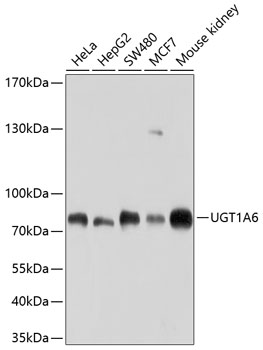
Figure 1. Western blot analysis of UGT1A6 using anti-UGT1A6 antibody (A02239-1). Electrophoresis was performed on a 5-20% SDS-PAGE gel at 70V (Stacking gel) / 90V (Resolving gel) for 2-3 hours. The sample well of each lane was loaded with 50ug of sample under reducing conditions. Lane 1: human HEK293 whole cell lysates. After Electrophoresis, proteins were transferred to a Nitrocellulose membrane at 150mA for 50-90 minutes. Blocked the membrane with 5% Non-fat Milk/ TBS for 1.5 hour at RT. The membrane was incubated with rabbit anti-UGT1A6 antigen affinity purified polyclonal antibody (Catalog # A02239-1) at 0.5 microg/mL overnight at 4°C, then washed with TBS-0.1%Tween 3 times with 5 minutes each and probed with a goat anti-rabbit IgG-HRP secondary antibody at a dilution of 1:10000 for 1.5 hour at RT. The signal is developed using an Enhanced Chemiluminescent detection (ECL) kit (Catalog # EK1002) with Tanon 5200 system. A specific band was detected for UGT1A6 at approximately 55-61KD. The expected band size for UGT1A6 is at 61KD.
. UGT1A6 was detected in immunocytochemical section of U2OS cells. Enzyme antigen retrieval was performed using IHC enzyme antigen retrieval reagent (AR0022) for 15 mins. The cells were blocked with 10% goat serum. And then incubated with 2microg/mL rabbit anti-UGT1A6 Antibody (A02239-1) overnight at 4°C. DyLight®488 Conjugated Goat Anti-Rabbit IgG (BA1127) was used as secondary antibody at 1:100 dilution and incubated for 30 minutes at 37°C. The section was counterstained with DAPI. Visualize using a fluorescence microscope and filter sets appropriate for the label used.")
Figure 1. Western blot analysis of UGT1A6 using anti-UGT1A6 antibody (A02239-1). Electrophoresis was performed on a 5-20% SDS-PAGE gel at 70V (Stacking gel) / 90V (Resolving gel) for 2-3 hours. The sample well of each lane was loaded with 50ug of sample under reducing conditions. Lane 1: human HEK293 whole cell lysates. After Electrophoresis, proteins were transferred to a Nitrocellulose membrane at 150mA for 50-90 minutes. Blocked the membrane with 5% Non-fat Milk/ TBS for 1.5 hour at RT. The membrane was incubated with rabbit anti-UGT1A6 antigen affinity purified polyclonal antibody (Catalog # A02239-1) at 0.5 microg/mL overnight at 4°C, then washed with TBS-0.1%Tween 3 times with 5 minutes each and probed with a goat anti-rabbit IgG-HRP secondary antibody at a dilution of 1:10000 for 1.5 hour at RT. The signal is developed using an Enhanced Chemiluminescent detection (ECL) kit (Catalog # EK1002) with Tanon 5200 system. A specific band was detected for UGT1A6 at approximately 55-61KD. The expected band size for UGT1A6 is at 61KD.
Anti-UGT1A6 Antibody Picoband(r)
A02239-1-CARRIER-FREE
ApplicationsImmunoFluorescence, Western Blot, ELISA, ImmunoCytoChemistry
Product group Antibodies
ReactivityHuman
TargetUGT1A6
Overview
- SupplierBoster Bio
- Product NameAnti-UGT1A6 Antibody Picoband(r)
- Delivery Days Customer9
- ApplicationsImmunoFluorescence, Western Blot, ELISA, ImmunoCytoChemistry
- CertificationResearch Use Only
- ClonalityPolyclonal
- Concentration500 ug/ml
- Gene ID54578
- Target nameUGT1A6
- Target descriptionUDP glucuronosyltransferase family 1 member A6
- Target synonymsGNT1, HLUGP, HLUGP1, UDPGT, UDPGT 1-6, UGT-1A, UGT-1C, UGT-1E, UGT-1F, UGT1, UGT1-01, UGT1-03, UGT1-05, UGT1-06, UGT1.1, UGT1.3, UGT1.5, UGT1.6, UGT1A, UGT1A1, UGT1A3, UGT1A5, UGT1A6S, UGT1C, UGT1E, UGT1F, hUG-BR1, UDP-glucuronosyltransferase 1A6, Bilirubin-specific UDPGT isozyme 1, UDP glucuronosyltransferase 1 family, polypeptide A6, UDP glycosyltransferase 1 family, polypeptide A6, UDP-glucuronosyltransferase 1 family polypeptide A6s, UDP-glucuronosyltransferase 1-1, UDP-glucuronosyltransferase 1-3, UDP-glucuronosyltransferase 1-5, UDP-glucuronosyltransferase 1-6, UDP-glucuronosyltransferase 1-A, UDP-glucuronosyltransferase 1-C, UDP-glucuronosyltransferase 1-E, UDP-glucuronosyltransferase 1-F, UDP-glucuronosyltransferase 1A1, UDP-glucuronosyltransferase 1A3, UDP-glucuronosyltransferase 1A5, phenol-metabolizing UDP-glucuronosyltransferase
- HostRabbit
- IsotypeIgG
- Protein IDP19224
- Protein NameUDP-glucuronosyltransferase 1A6
- Scientific DescriptionBoster Bio Anti-UGT1A6 Antibody Picoband® catalog # A02239-1. Tested in ELISA, IF, ICC, WB applications. This antibody reacts with Human. The brand Picoband indicates this is a premium antibody that guarantees superior quality, high affinity, and strong signals with minimal background in Western blot applications. Only our best-performing antibodies are designated as Picoband, ensuring unmatched performance.
- ReactivityHuman
- Storage Instruction-20°C,2°C to 8°C
- UNSPSC12352203
Related products
Product group Antibodies
Anti-UGT1A6 Antibody144-10033
ApplicationsWestern Blot, ImmunoHistoChemistry
ReactivityHuman, Mouse, Rat
TargetUGT1A6
- SizePrice
Product group Antibodies
UGT1A6 antibodyGTX113980
ApplicationsWestern Blot, ImmunoHistoChemistry, ImmunoHistoChemistry Paraffin
ReactivityHuman, Mouse
TargetUGT1A6
- SizePrice
Product group Antibodies
Anti-UGT1A6 AntibodyA11219
ApplicationsWestern Blot
ReactivityHuman, Mouse, Rat
- SizePrice
Product group Antibodies

UGT1A6 AntibodyLS-C496745
ApplicationsWestern Blot, ImmunoHistoChemistry
ReactivityHuman, Mouse
TargetUGT1A6
- SizePrice
Product group Antibodies
UGT1A6 AntibodyCSB-PA025579LA01HU
ApplicationsELISA, ImmunoHistoChemistry
ReactivityHuman
TargetUGT1A6
- SizePrice

Product group Antibodies
Anti-UGT1A6 AntibodyHPA054065
ApplicationsImmunoHistoChemistry
ReactivityHuman
TargetUGT1A6
- SizePrice