

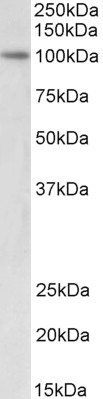
Anti-UHRF1 Antibody
A83277
ApplicationsWestern Blot, ELISA
Product group Antibodies
ReactivityHuman
Overview
- SupplierAntibodies.com
- Product NameAnti-UHRF1 Antibody
- Delivery Days Customer7
- ApplicationsWestern Blot, ELISA
- CertificationResearch Use Only
- ClonalityPolyclonal
- Concentration500 ug/ml
- ConjugateUnconjugated
- HostGoat
- IsotypeIgG
- Scientific DescriptionGoat polyclonal antibody to UHRF1.
- ReactivityHuman
- UNSPSC12352203
Related products

Product group Antibodies
Anti-UHRF1 Antibody Picoband(r)A01156-1-CARRIER-FREE
ApplicationsFlow Cytometry, ImmunoFluorescence, Western Blot, ELISA, ImmunoCytoChemistry
ReactivityHuman, Mouse
TargetUHRF1
- SizePrice
Product group Antibodies
Anti-UHRF1 Antibody144-02343
ApplicationsImmunoFluorescence, Western Blot, ImmunoHistoChemistry
ReactivityHuman
TargetUHRF1
- SizePrice
Product group Antibodies

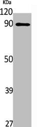
UHRF1 AntibodyLS-C747684
ApplicationsWestern Blot
ReactivityHuman, Mouse
TargetUHRF1
- SizePrice
Product group Antibodies
UHRF1 Polyclonal AntibodyBS-25668R
ApplicationsImmunoFluorescence, ImmunoHistoChemistry, ImmunoHistoChemistry Frozen, ImmunoHistoChemistry Paraffin
ReactivityCanine, Human, Mouse, Rat
TargetUHRF1
- SizePrice
Product group Antibodies
UHRF1 AntibodyCSB-PA004690
ApplicationsWestern Blot, ELISA
ReactivityHuman
TargetUHRF1
- SizePrice

Product group Antibodies
Anti-UHRF1 AntibodyHPA049408
ApplicationsImmunoCytoChemistry, ImmunoHistoChemistry
ReactivityHuman
TargetUHRF1
- SizePrice
Product group Antibodies
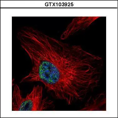
UHRF1 antibodyGTX103925
ApplicationsImmunoFluorescence, Western Blot, ImmunoCytoChemistry, ImmunoHistoChemistry, ImmunoHistoChemistry Paraffin
ReactivityHuman
TargetUHRF1
- SizePrice