


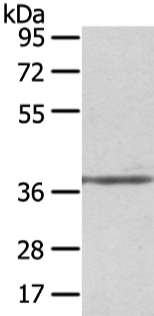
Figure 1. Western blot analysis of Unrip using anti-Unrip antibody (PB9407). Electrophoresis was performed on a 5-20% SDS-PAGE gel at 70V (Stacking gel) / 90V (Resolving gel) for 2-3 hours. The sample well of each lane was loaded with 50ug of sample under reducing conditions. Lane 1: human HEK293 whole cell lysates Lane 2: human Jurkat whole cell lysates Lane 3: human PC-3 whole cell lysates Lane 4: human THP-1 whole cell lysates Lane 5: rat brain tissue lysates Lane 6: mouse HEPA1-6 whole cell lysates Lane 7: mouse RAW246.7 whole cell lysates After Electrophoresis, proteins were transferred to a Nitrocellulose membrane at 150mA for 50-90 minutes. Blocked the membrane with 5% Non-fat Milk/ TBS for 1.5 hour at RT. The membrane was incubated with rabbit anti-Unrip antigen affinity purified polyclonal antibody (Catalog # PB9407) at 0.5 microg/mL overnight at 4°C, then washed with TBS-0.1%Tween 3 times with 5 minutes each and probed with a goat anti-rabbit IgG-HRP secondary antibody at a dilution of 1:10000 for 1.5 hour at RT. The signal is developed using an Enhanced Chemiluminescent detection (ECL) kit (Catalog # EK1002) with Tanon 5200 system. A specific band was detected for Unrip at approximately 39KD. The expected band size for Unrip is at 39KD.
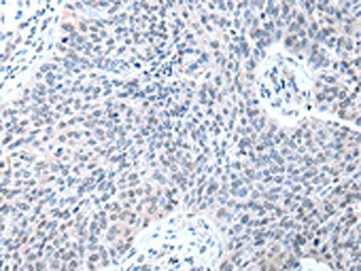
. Unrip was detected in a paraffin-embedded section of human mammary cancer tissue. Heat mediated antigen retrieval was performed in EDTA buffer (pH 8.0, epitope retrieval solution). The tissue section was blocked with 10% goat serum. The tissue section was then incubated with 1 microg/ml rabbit anti-Unrip Antibody (PB9407) overnight at 4°C. Biotinylated goat anti-rabbit IgG was used as secondary antibody and incubated for 30 minutes at 37°C. The tissue section was developed using Strepavidin-Biotin-Complex (SABC) (Catalog # SA1022) with DAB as the chromogen.")
Figure 1. Western blot analysis of Unrip using anti-Unrip antibody (PB9407). Electrophoresis was performed on a 5-20% SDS-PAGE gel at 70V (Stacking gel) / 90V (Resolving gel) for 2-3 hours. The sample well of each lane was loaded with 50ug of sample under reducing conditions. Lane 1: human HEK293 whole cell lysates Lane 2: human Jurkat whole cell lysates Lane 3: human PC-3 whole cell lysates Lane 4: human THP-1 whole cell lysates Lane 5: rat brain tissue lysates Lane 6: mouse HEPA1-6 whole cell lysates Lane 7: mouse RAW246.7 whole cell lysates After Electrophoresis, proteins were transferred to a Nitrocellulose membrane at 150mA for 50-90 minutes. Blocked the membrane with 5% Non-fat Milk/ TBS for 1.5 hour at RT. The membrane was incubated with rabbit anti-Unrip antigen affinity purified polyclonal antibody (Catalog # PB9407) at 0.5 microg/mL overnight at 4°C, then washed with TBS-0.1%Tween 3 times with 5 minutes each and probed with a goat anti-rabbit IgG-HRP secondary antibody at a dilution of 1:10000 for 1.5 hour at RT. The signal is developed using an Enhanced Chemiluminescent detection (ECL) kit (Catalog # EK1002) with Tanon 5200 system. A specific band was detected for Unrip at approximately 39KD. The expected band size for Unrip is at 39KD.
Anti-Unrip/STRAP Antibody Picoband(r)
PB9407-BIOTIN
ApplicationsWestern Blot, ImmunoHistoChemistry
Product group Antibodies
ReactivityHamster, Human, Mouse, Rat
TargetSTRAP
Overview
- SupplierBoster Bio
- Product NameAnti-Unrip/STRAP Antibody Picoband(r)
- Delivery Days Customer9
- Application Supplier NoteWB: The detection limit for Unrip is approximately 0.1ng/lane under reducing conditions. Tested Species: In-house tested species with positive results. By Heat: Boiling the paraffin sections in 10mM citrate buffer, pH6.0, for 20mins is required for the staining of formalin/paraffin sections. Other applications have not been tested. Optimal dilutions should be determined by end users.
- ApplicationsWestern Blot, ImmunoHistoChemistry
- CertificationResearch Use Only
- ClonalityPolyclonal
- Concentration500 ug/ml
- ConjugateBiotin
- Gene ID11171
- Target nameSTRAP
- Target descriptionserine/threonine kinase receptor associated protein
- Target synonymsMAWD, PT-WD, UNRIP, serine-threonine kinase receptor-associated protein, MAP activator with WD repeats, MAPK activator with WD repeats, WD-40 repeat protein PT-WD, mitogen-activated protein kinase activator with WD repeats, unr-interacting protein
- HostRabbit
- IsotypeIgG
- Protein IDQ9Y3F4
- Protein NameSerine-threonine kinase receptor-associated protein
- Scientific DescriptionBoster Bio Anti-Unrip/STRAP Antibody Picoband® catalog # PB9407. Tested in IHC, WB applications. This antibody reacts with Human, Mouse, Rat. The brand Picoband indicates this is a premium antibody that guarantees superior quality, high affinity, and strong signals with minimal background in Western blot applications. Only our best-performing antibodies are designated as Picoband, ensuring unmatched performance.
- ReactivityHamster, Human, Mouse, Rat
- Storage Instruction-20°C,2°C to 8°C
- UNSPSC12352203
Related products
Product group Antibodies
Unrip Polyclonal AntibodyBS-5132R
ApplicationsImmunoFluorescence, Western Blot, ELISA, ImmunoCytoChemistry, ImmunoHistoChemistry, ImmunoHistoChemistry Frozen, ImmunoHistoChemistry Paraffin
ReactivityBovine, Equine, Human, Mouse, Porcine, Rabbit, Rat
TargetSTRAP
- SizePrice
Product group Antibodies
Anti-STRAP AntibodyA38230
ApplicationsWestern Blot, ImmunoHistoChemistry
ReactivityHuman, Mouse
- SizePrice
Product group Antibodies
Anti-STRAP Antibody144-05964
ApplicationsWestern Blot
ReactivityHuman, Mouse, Rat
TargetSTRAP
- SizePrice
Product group Antibodies
STRAP antibody [N1C3]GTX118603
ApplicationsImmunoFluorescence, Western Blot, ImmunoCytoChemistry, ImmunoHistoChemistry, ImmunoHistoChemistry Paraffin
ReactivityHuman, Mouse, Rat
TargetSTRAP
- SizePrice
Product group Antibodies

STRAP / MAWD AntibodyLS-C334420
ApplicationsWestern Blot
ReactivityHuman, Mouse, Rat
TargetSTRAP
- SizePrice

Product group Antibodies
Anti-STRAP AntibodyHPA027320
ApplicationsWestern Blot, ImmunoHistoChemistry
ReactivityHuman, Mouse, Rat
TargetSTRAP
- SizePrice
Product group Antibodies
STRAP AntibodyCSB-PA227440
ApplicationsELISA, ImmunoHistoChemistry
ReactivityHuman, Mouse, Rat
TargetSTRAP
- SizePrice
Product group Antibodies
Anti-Unrip/STRAP Antibody Picoband(r)PB9407-CARRIER-FREE
ApplicationsWestern Blot, ImmunoHistoChemistry
ReactivityHamster, Human, Mouse, Rat
TargetSTRAP
- SizePrice