


Immunofluorescent staining of human cell line Hep G2 shows localization to nucleus & vesicles.
Immunofluorescent staining of human cell line Hep G2 shows localization to nucleus & vesicles.
Anti-UPF3A Antibody
HPA061316
ApplicationsImmunoCytoChemistry
Product group Antibodies
ReactivityHuman
TargetUPF3A
Overview
- SupplierAtlas Antibodies
- Product NameAnti-UPF3A Antibody
- Delivery Days Customer4
- ApplicationsImmunoCytoChemistry
- CertificationResearch Use Only
- ClonalityPolyclonal
- ConjugateUnconjugated
- Gene ID65110
- Target nameUPF3A
- Target descriptionUPF3A regulator of nonsense mediated mRNA decay
- Target synonymsHUPF3A, RENT3A, UPF3, regulator of nonsense transcripts 3A, UPF3 regulator of nonsense transcripts homolog A, hUpf3, nonsense mRNA reducing factor 3A, up-frameshift suppressor 3 homolog A
- HostRabbit
- IsotypeIgG
- Protein IDQ9H1J1
- Protein NameRegulator of nonsense transcripts 3A
- Scientific DescriptionRecombinant Protein Epitope Signature Tag (PrEST) antigen sequence
- ReactivityHuman
- Storage Instruction-20°C,2°C to 8°C
- UNSPSC41116161
Datasheet
MSDS
Related products
Product group Antibodies
Anti-UPF3A Antibody144-61267
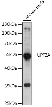
ApplicationsWestern Blot
ReactivityHuman, Mouse
TargetUPF3A
- SizePrice
Product group Antibodies
Anti-UPF3A AntibodyA90511
ApplicationsImmunoFluorescence, Western Blot, ImmunoCytoChemistry
ReactivityHuman, Mouse, Rat
- SizePrice
Product group Antibodies

UPF3A / UPF3 AntibodyLS-C833619
ApplicationsImmunoFluorescence
ReactivityHuman, Mouse, Rat
TargetUPF3A
- SizePrice
Product group Antibodies
UPF3A AntibodyCSB-PA025652GA01HU
ApplicationsWestern Blot, ELISA
ReactivityHuman, Mouse, Rat
TargetUPF3A
- SizePrice
Product group Antibodies

ApplicationsELISA
ReactivityBovine, Human
TargetUPF3A
- SizePrice

Product group Antibodies
Anti-UPF3A AntibodyHPA018325
ApplicationsImmunoHistoChemistry
ReactivityHuman
TargetUPF3A
- SizePrice
Product group Antibodies
Anti-UPF3A AntibodyHPA018325
ApplicationsImmunoHistoChemistry
ReactivityHuman
TargetUPF3A
- SizePrice