


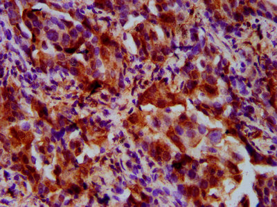
Immunohistochemical staining of human placenta shows strong cytoplasmic positivity in trophoblastic cells.
. Lane 2: NBT-II cell lysate (Rat Wistar bladder tumour cells). Lane 3: PC12 cell lysate (Pheochromocytoma of rat adrenal medulla)")
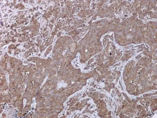
Immunohistochemical staining of human placenta shows strong cytoplasmic positivity in trophoblastic cells.
Anti-UPF3B Antibody
HPA001800
ApplicationsWestern Blot, ImmunoHistoChemistry
Product group Antibodies
ReactivityHuman, Mouse, Rat
TargetUPF3B
Overview
- SupplierAtlas Antibodies
- Product NameAnti-UPF3B Antibody
- Delivery Days Customer4
- ApplicationsWestern Blot, ImmunoHistoChemistry
- CertificationResearch Use Only
- ClonalityPolyclonal
- ConjugateUnconjugated
- Gene ID65109
- Target nameUPF3B
- Target descriptionUPF3B regulator of nonsense mediated mRNA decay
- Target synonymsHUPF3B, MRX62, MRX82, MRXS14, RENT3B, UPF3BP1, UPF3BP2, UPF3BP3, UPF3X, Upf3p-X, regulator of nonsense transcripts 3B, UPF3 regulator of nonsense transcripts homolog B, nonsense mRNA reducing factor 3B, up-frameshift suppressor 3 homolog B, up-frameshift suppressor 3 homolog on chromosome X
- HostRabbit
- IsotypeIgG
- Protein IDQ9BZI7
- Protein NameRegulator of nonsense transcripts 3B
- Scientific DescriptionRecombinant Protein Epitope Signature Tag (PrEST) antigen sequence
- ReactivityHuman, Mouse, Rat
- Storage Instruction-20°C,2°C to 8°C
- UNSPSC41116161
Datasheet
MSDS
Related products
Product group Antibodies

UPF3B AntibodyLS-C750156
ApplicationsWestern Blot
ReactivityHuman, Mouse
TargetUPF3B
- SizePrice

Product group Antibodies
Anti-UPF3B AntibodyHPA001592
ApplicationsWestern Blot, ImmunoCytoChemistry
ReactivityHuman
TargetUPF3B
- SizePrice

Product group Antibodies
Anti-UPF3B-25ulHPA001882
ApplicationsWestern Blot, ImmunoCytoChemistry, ImmunoHistoChemistry
ReactivityHuman
- SizePrice
Product group Antibodies
UPF3B AntibodyCSB-PA883646LA01HU
ApplicationsImmunoFluorescence, ELISA, ImmunoHistoChemistry
ReactivityHuman
TargetUPF3B
- SizePrice

Product group Antibodies
Anti-UPF3B/RENT3B Antibody Picoband(r)PB9843-CARRIER-FREE
ApplicationsWestern Blot, ImmunoHistoChemistry
ReactivityBovine, Human, Mouse, Rat
TargetUPF3B
- SizePrice
Product group Antibodies
UPF3B antibodyGTX130493
ApplicationsWestern Blot, ImmunoHistoChemistry, ImmunoHistoChemistry Paraffin
ReactivityHuman
TargetUPF3B
- SizePrice