

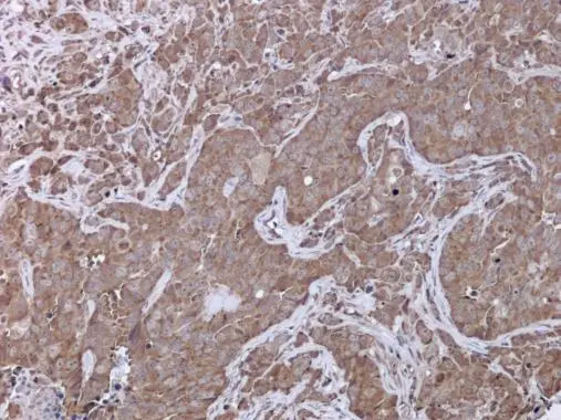
UPF3B antibody detects UPF3B protein at cytoplasm in AsPC-1 xenograft by immunohistochemical analysis. Sample: Paraffin-embedded AsPC-1 xenograft. UPF3B antibody (GTX130493) diluted at 1:500.
Antigen Retrieval: Citrate buffer, pH 6.0, 15 min
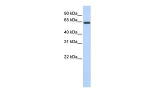
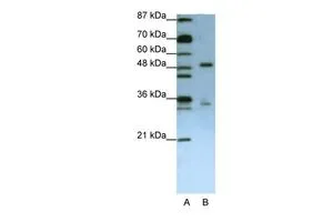
 were separated by 10% SDS-PAGE, and the membrane was blotted with UPF3B antibody (GTX130493) diluted at 1:1000.")
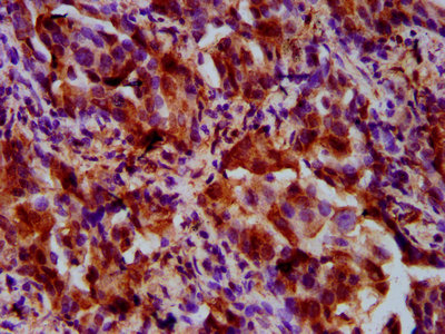
UPF3B antibody detects UPF3B protein at cytoplasm in AsPC-1 xenograft by immunohistochemical analysis. Sample: Paraffin-embedded AsPC-1 xenograft. UPF3B antibody (GTX130493) diluted at 1:500.
Antigen Retrieval: Citrate buffer, pH 6.0, 15 min
UPF3B antibody
GTX130493
ApplicationsWestern Blot, ImmunoHistoChemistry, ImmunoHistoChemistry Paraffin
Product group Antibodies
ReactivityHuman
TargetUPF3B
Overview
- SupplierGeneTex
- Product NameUPF3B antibody
- Delivery Days Customer9
- Application Supplier NoteWB: 1:500-1:3000. IHC-P: 1:100-1:1000. *Optimal dilutions/concentrations should be determined by the researcher.Not tested in other applications.
- ApplicationsWestern Blot, ImmunoHistoChemistry, ImmunoHistoChemistry Paraffin
- CertificationResearch Use Only
- ClonalityPolyclonal
- Concentration1.27 mg/ml
- ConjugateUnconjugated
- Gene ID65109
- Target nameUPF3B
- Target descriptionUPF3B regulator of nonsense mediated mRNA decay
- Target synonymsHUPF3B, MRX62, MRX82, MRXS14, RENT3B, UPF3BP1, UPF3BP2, UPF3BP3, UPF3X, Upf3p-X, regulator of nonsense transcripts 3B, UPF3 regulator of nonsense transcripts homolog B, nonsense mRNA reducing factor 3B, up-frameshift suppressor 3 homolog B, up-frameshift suppressor 3 homolog on chromosome X
- HostRabbit
- IsotypeIgG
- Protein IDQ9BZI7
- Protein NameRegulator of nonsense transcripts 3B
- Scientific DescriptionThis gene encodes a protein that is part of a post-splicing multiprotein complex involved in both mRNA nuclear export and mRNA surveillance. The encoded protein is one of two functional homologs to yeast Upf3p. mRNA surveillance detects exported mRNAs with truncated open reading frames and initiates nonsense-mediated mRNA decay (NMD). When translation ends upstream from the last exon-exon junction, this triggers NMD to degrade mRNAs containing premature stop codons. This protein binds to the mRNA and remains bound after nuclear export, acting as a nucleocytoplasmic shuttling protein. It forms with Y14 a complex that binds specifically 20 nt upstream of exon-exon junctions. This gene is located on the long arm of chromosome X. Two splice variants encoding different isoforms have been found for this gene. [provided by RefSeq]
- ReactivityHuman
- Storage Instruction-20°C or -80°C,2°C to 8°C
- UNSPSC41116161
Datasheet
Related products
Product group Antibodies

UPF3B AntibodyLS-C750156
ApplicationsWestern Blot
ReactivityHuman, Mouse
TargetUPF3B
- SizePrice

Product group Antibodies
Anti-UPF3B AntibodyHPA001800
ApplicationsWestern Blot, ImmunoHistoChemistry
ReactivityHuman, Mouse, Rat
TargetUPF3B
- SizePrice
Product group Antibodies
UPF3B AntibodyCSB-PA883646LA01HU
ApplicationsImmunoFluorescence, ELISA, ImmunoHistoChemistry
ReactivityHuman
TargetUPF3B
- SizePrice

Product group Antibodies
Anti-UPF3B/RENT3B Antibody Picoband(r)PB9843-CARRIER-FREE
ApplicationsWestern Blot, ImmunoHistoChemistry
ReactivityBovine, Human, Mouse, Rat
TargetUPF3B
- SizePrice
Product group Antibodies
UPF3B antibody, InternalGTX47228
ApplicationsWestern Blot
ReactivityHuman
TargetUPF3B
- SizePrice
Product group Antibodies
UPF3B antibody, N-termGTX47258
ApplicationsWestern Blot, ImmunoHistoChemistry, ImmunoHistoChemistry Paraffin
ReactivityHuman
TargetUPF3B
- SizePrice