


Figure 1. Western blot analysis of UPF3B/RENT3B using anti-UPF3B/RENT3B antibody (PB9843). Electrophoresis was performed on a 5-20% SDS-PAGE gel at 70V (Stacking gel) / 90V (Resolving gel) for 2-3 hours. The sample well of each lane was loaded with 30 ug of sample under reducing conditions. Lane 1: human Hela whole cell lysates, Lane 2: human 22RV1 whole cell lysates, Lane 3: human U87 whole cell lysates, Lane 4: human Jurkat whole cell lysates, Lane 5: rat brain tissue lysates. After electrophoresis, proteins were transferred to a nitrocellulose membrane at 150 mA for 50-90 minutes. Blocked the membrane with 5% non-fat milk/TBS for 1.5 hour at RT. The membrane was incubated with rabbit anti-UPF3B/RENT3B antigen affinity purified polyclonal antibody (Catalog # PB9843) at 0.5 microg/mL overnight at 4°C, then washed with TBS-0.1%Tween 3 times with 5 minutes each and probed with a goat anti-rabbit IgG-HRP secondary antibody at a dilution of 1:5000 for 1.5 hour at RT. The signal is developed using an Enhanced Chemiluminescent detection (ECL) kit (Catalog # EK1002) with Tanon 5200 system. A specific band was detected for UPF3B/RENT3B at approximately 58 kDa. The expected band size for UPF3B/RENT3B is at 58 kDa.
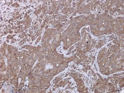
. UPF3B/RENT3B was detected in a paraffin-embedded section of mouse testis tissue. Heat mediated antigen retrieval was performed in EDTA buffer (pH 8.0, epitope retrieval solution). The tissue section was blocked with 10% goat serum. The tissue section was then incubated with 1 microg/ml rabbit anti-UPF3B/RENT3B Antibody (PB9843) overnight at 4°C. Biotinylated goat anti-rabbit IgG was used as secondary antibody and incubated for 30 minutes at 37°C. The tissue section was developed using Strepavidin-Biotin-Complex (SABC) (Catalog # SA1022) with DAB as the chromogen.")
. UPF3B/RENT3B was detected in a paraffin-embedded section of rat testis tissue. Heat mediated antigen retrieval was performed in EDTA buffer (pH 8.0, epitope retrieval solution). The tissue section was blocked with 10% goat serum. The tissue section was then incubated with 1 microg/ml rabbit anti-UPF3B/RENT3B Antibody (PB9843) overnight at 4°C. Biotinylated goat anti-rabbit IgG was used as secondary antibody and incubated for 30 minutes at 37°C. The tissue section was developed using Strepavidin-Biotin-Complex (SABC) (Catalog # SA1022) with DAB as the chromogen.")
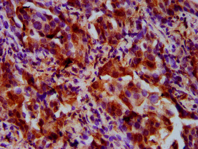
. UPF3B/RENT3B was detected in a paraffin-embedded section of human placenta tissue. Heat mediated antigen retrieval was performed in EDTA buffer (pH 8.0, epitope retrieval solution). The tissue section was blocked with 10% goat serum. The tissue section was then incubated with 1 microg/ml rabbit anti-UPF3B/RENT3B Antibody (PB9843) overnight at 4°C. Biotinylated goat anti-rabbit IgG was used as secondary antibody and incubated for 30 minutes at 37°C. The tissue section was developed using Strepavidin-Biotin-Complex (SABC) (Catalog # SA1022) with DAB as the chromogen.")
Figure 1. Western blot analysis of UPF3B/RENT3B using anti-UPF3B/RENT3B antibody (PB9843). Electrophoresis was performed on a 5-20% SDS-PAGE gel at 70V (Stacking gel) / 90V (Resolving gel) for 2-3 hours. The sample well of each lane was loaded with 30 ug of sample under reducing conditions. Lane 1: human Hela whole cell lysates, Lane 2: human 22RV1 whole cell lysates, Lane 3: human U87 whole cell lysates, Lane 4: human Jurkat whole cell lysates, Lane 5: rat brain tissue lysates. After electrophoresis, proteins were transferred to a nitrocellulose membrane at 150 mA for 50-90 minutes. Blocked the membrane with 5% non-fat milk/TBS for 1.5 hour at RT. The membrane was incubated with rabbit anti-UPF3B/RENT3B antigen affinity purified polyclonal antibody (Catalog # PB9843) at 0.5 microg/mL overnight at 4°C, then washed with TBS-0.1%Tween 3 times with 5 minutes each and probed with a goat anti-rabbit IgG-HRP secondary antibody at a dilution of 1:5000 for 1.5 hour at RT. The signal is developed using an Enhanced Chemiluminescent detection (ECL) kit (Catalog # EK1002) with Tanon 5200 system. A specific band was detected for UPF3B/RENT3B at approximately 58 kDa. The expected band size for UPF3B/RENT3B is at 58 kDa.
Anti-UPF3B/RENT3B Antibody Picoband(r)
PB9843-CARRIER-FREE
ApplicationsWestern Blot, ImmunoHistoChemistry
Product group Antibodies
ReactivityBovine, Human, Mouse, Rat
TargetUPF3B
Overview
- SupplierBoster Bio
- Product NameAnti-UPF3B/RENT3B Antibody Picoband(r)
- Delivery Days Customer9
- Application Supplier NoteTested Species: In-house tested species with positive results. By Heat: Boiling the paraffin sections in 10mM citrate buffer, pH6.0, for 20mins is required for the staining of formalin/paraffin sections. Other applications have not been tested. Optimal dilutions should be determined by end users.
- ApplicationsWestern Blot, ImmunoHistoChemistry
- CertificationResearch Use Only
- ClonalityPolyclonal
- Concentration500 ug/ml
- Gene ID65109
- Target nameUPF3B
- Target descriptionUPF3B regulator of nonsense mediated mRNA decay
- Target synonymsHUPF3B, MRX62, MRX82, MRXS14, RENT3B, UPF3BP1, UPF3BP2, UPF3BP3, UPF3X, Upf3p-X, regulator of nonsense transcripts 3B, UPF3 regulator of nonsense transcripts homolog B, nonsense mRNA reducing factor 3B, up-frameshift suppressor 3 homolog B, up-frameshift suppressor 3 homolog on chromosome X
- HostRabbit
- IsotypeIgG
- Protein IDQ9BZI7
- Protein NameRegulator of nonsense transcripts 3B
- Scientific DescriptionBoster Bio Anti-UPF3B/RENT3B Antibody Picoband® catalog # PB9843. Tested in IHC, WB applications. This antibody reacts with Human, Mouse, Rat. The brand Picoband indicates this is a premium antibody that guarantees superior quality, high affinity, and strong signals with minimal background in Western blot applications. Only our best-performing antibodies are designated as Picoband, ensuring unmatched performance.
- ReactivityBovine, Human, Mouse, Rat
- Storage Instruction-20°C,2°C to 8°C
- UNSPSC12352203
Related products
Product group Antibodies

UPF3B AntibodyLS-C750156
ApplicationsWestern Blot
ReactivityHuman, Mouse
TargetUPF3B
- SizePrice

Product group Antibodies
Anti-UPF3B AntibodyHPA001800
ApplicationsWestern Blot, ImmunoHistoChemistry
ReactivityHuman, Mouse, Rat
TargetUPF3B
- SizePrice
Product group Antibodies
UPF3B AntibodyCSB-PA883646LA01HU
ApplicationsImmunoFluorescence, ELISA, ImmunoHistoChemistry
ReactivityHuman
TargetUPF3B
- SizePrice
Product group Antibodies
UPF3B antibodyGTX130493
ApplicationsWestern Blot, ImmunoHistoChemistry, ImmunoHistoChemistry Paraffin
ReactivityHuman
TargetUPF3B
- SizePrice