


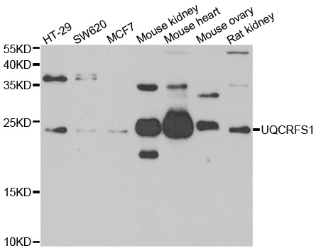
Figure 1. Western blot analysis of UQCRFS1 using anti-UQCRFS1 antibody (M08781-1). Electrophoresis was performed on a 5-20% SDS-PAGE gel at 70V (Stacking gel) / 90V (Resolving gel) for 2-3 hours. The sample well of each lane was loaded with 30 ug of sample under reducing conditions. Lane 1: human Raji whole cell lysates, Lane 2: human A549 whole cell lysates, Lane 3: human 293T whole cell lysates, Lane 4: human RT4 whole cell lysates, Lane 5: rat liver tissue lysates, Lane 6: mouse ovary tissue lysates, Lane 7: mouse liver tissue lysates. After electrophoresis, proteins were transferred to a nitrocellulose membrane at 150 mA for 50-90 minutes. Blocked the membrane with 5% non-fat milk/TBS for 1.5 hour at RT. The membrane was incubated with rabbit anti-UQCRFS1 antigen affinity purified monoclonal antibody (Catalog # M08781-1) at 1:500 overnight at 4°C, then washed with TBS-0.1%Tween 3 times with 5 minutes each and probed with a goat anti-rabbit IgG-HRP secondary antibody at a dilution of 1:500 for 1.5 hour at RT. The signal is developed using an Enhanced Chemiluminescent detection (ECL) kit (Catalog # EK1002) with Tanon 5200 system. A specific band was detected for UQCRFS1 at approximately 23 kDa. The expected band size for UQCRFS1 is at 30 kDa.
. UQCRFS1 was detected in a paraffin-embedded section of mouse heart tissue. Heat mediated antigen retrieval was performed in EDTA buffer (pH 8.0, epitope retrieval solution). The tissue section was blocked with 10% goat serum. The tissue section was then incubated with 1:100 rabbit anti-UQCRFS1 Antibody (M08781-1) overnight at 4°C. Peroxidase Conjugated Goat Anti-rabbit IgG was used as secondary antibody and incubated for 30 minutes at 37°C. The tissue section was developed using HRP Conjugated Rabbit IgG Super Vision Assay Kit (Catalog # SV0002) with DAB as the chromogen.")
. UQCRFS1 was detected in a paraffin-embedded section of mouse heart tissue. Heat mediated antigen retrieval was performed in EDTA buffer (pH 8.0, epitope retrieval solution). The tissue section was blocked with 10% goat serum. The tissue section was then incubated with 1:100 rabbit anti-UQCRFS1 Antibody (M08781-1) overnight at 4°C. Peroxidase Conjugated Goat Anti-rabbit IgG was used as secondary antibody and incubated for 30 minutes at 37°C. The tissue section was developed using HRP Conjugated Rabbit IgG Super Vision Assay Kit (Catalog # SV0002) with DAB as the chromogen.")
. UQCRFS1 was detected in a paraffin-embedded section of rat heart tissue. Heat mediated antigen retrieval was performed in EDTA buffer (pH 8.0, epitope retrieval solution). The tissue section was blocked with 10% goat serum. The tissue section was then incubated with 1:100 rabbit anti-UQCRFS1 Antibody (M08781-1) overnight at 4°C. Peroxidase Conjugated Goat Anti-rabbit IgG was used as secondary antibody and incubated for 30 minutes at 37°C. The tissue section was developed using HRP Conjugated Rabbit IgG Super Vision Assay Kit (Catalog # SV0002) with DAB as the chromogen.")
. UQCRFS1 was detected in a paraffin-embedded section of rat heart tissue. Heat mediated antigen retrieval was performed in EDTA buffer (pH 8.0, epitope retrieval solution). The tissue section was blocked with 10% goat serum. The tissue section was then incubated with 1:100 rabbit anti-UQCRFS1 Antibody (M08781-1) overnight at 4°C. Peroxidase Conjugated Goat Anti-rabbit IgG was used as secondary antibody and incubated for 30 minutes at 37°C. The tissue section was developed using HRP Conjugated Rabbit IgG Super Vision Assay Kit (Catalog # SV0002) with DAB as the chromogen.")
Figure 1. Western blot analysis of UQCRFS1 using anti-UQCRFS1 antibody (M08781-1). Electrophoresis was performed on a 5-20% SDS-PAGE gel at 70V (Stacking gel) / 90V (Resolving gel) for 2-3 hours. The sample well of each lane was loaded with 30 ug of sample under reducing conditions. Lane 1: human Raji whole cell lysates, Lane 2: human A549 whole cell lysates, Lane 3: human 293T whole cell lysates, Lane 4: human RT4 whole cell lysates, Lane 5: rat liver tissue lysates, Lane 6: mouse ovary tissue lysates, Lane 7: mouse liver tissue lysates. After electrophoresis, proteins were transferred to a nitrocellulose membrane at 150 mA for 50-90 minutes. Blocked the membrane with 5% non-fat milk/TBS for 1.5 hour at RT. The membrane was incubated with rabbit anti-UQCRFS1 antigen affinity purified monoclonal antibody (Catalog # M08781-1) at 1:500 overnight at 4°C, then washed with TBS-0.1%Tween 3 times with 5 minutes each and probed with a goat anti-rabbit IgG-HRP secondary antibody at a dilution of 1:500 for 1.5 hour at RT. The signal is developed using an Enhanced Chemiluminescent detection (ECL) kit (Catalog # EK1002) with Tanon 5200 system. A specific band was detected for UQCRFS1 at approximately 23 kDa. The expected band size for UQCRFS1 is at 30 kDa.
Anti-UQCRFS1 Rabbit Monoclonal Antibody
M08781-1
ApplicationsFlow Cytometry, ImmunoFluorescence, ImmunoPrecipitation, Western Blot, ImmunoCytoChemistry, ImmunoHistoChemistry
Product group Antibodies
ReactivityHuman, Mouse, Rat
TargetUQCRFS1
Overview
- SupplierBoster Bio
- Product NameAnti-UQCRFS1 Rabbit Monoclonal Antibody
- Delivery Days Customer9
- ApplicationsFlow Cytometry, ImmunoFluorescence, ImmunoPrecipitation, Western Blot, ImmunoCytoChemistry, ImmunoHistoChemistry
- CertificationResearch Use Only
- ClonalityMonoclonal
- Clone ID19U18
- Gene ID7386
- Target nameUQCRFS1
- Target descriptionubiquinol-cytochrome c reductase, Rieske iron-sulfur polypeptide 1
- Target synonymsMC3DN10, RIP1, RIS1, RISP, UQCR5, cytochrome b-c1 complex subunit Rieske, mitochondrial, Rieske iron-sulfur protein, Rieske protein UQCRFS1, complex III subunit 5, cytochrome b-c1 complex subunit 5, epididymis secretory sperm binding protein, ubiquinol-cytochrome c reductase iron-sulfur subunit
- HostRabbit
- IsotypeIgG
- Protein IDP47985
- Protein NameCytochrome b-c1 complex subunit Rieske, mitochondrial
- Scientific DescriptionBoster Bio Anti-UQCRFS1 Rabbit Monoclonal Antibody catalog # M08781-1. Tested in WB, IHC, ICC/IF, IP, Flow Cytometry applications. This antibody reacts with Human, Mouse, Rat.
- ReactivityHuman, Mouse, Rat
- Storage Instruction-20°C
- UNSPSC12352203
Related products
Product group Antibodies
Anti-UQCRFS1 AntibodyA31603
ApplicationsWestern Blot, ImmunoHistoChemistry
ReactivityHuman, Mouse, Rat
- SizePrice
Product group Antibodies

UQCRFS1 AntibodyLS-C749084
ApplicationsWestern Blot, ImmunoHistoChemistry
ReactivityHuman, Mouse
TargetUQCRFS1
- SizePrice
Product group Antibodies
UQCRFS1 AntibodyCSB-PA04645A0RB
ApplicationsWestern Blot, ELISA, ImmunoHistoChemistry
ReactivityHuman
TargetUQCRFS1
- SizePrice

Product group Antibodies
Anti-UQCRFS1 AntibodyHPA041863
ApplicationsWestern Blot, ImmunoCytoChemistry, ImmunoHistoChemistry
ReactivityHuman, Rat
TargetUQCRFS1
- SizePrice
Product group Antibodies
UQCRFS1 Polyclonal AntibodyCAC14065
ApplicationsWestern Blot, ELISA, ImmunoHistoChemistry
TargetUQCRFS1
- SizePrice
Product group Antibodies
UQCRFS1 antibody [N1C3]GTX102150
ApplicationsImmunoFluorescence, Western Blot, ImmunoCytoChemistry
ReactivityHuman, Mouse
TargetUQCRFS1
- SizePrice
Product group Antibodies
Anti-UQCRFS1 Antibody144-06784
ApplicationsImmunoFluorescence, Western Blot
ReactivityHuman, Mouse, Rat
TargetUQCRFS1
- SizePrice
Product group Antibodies
UQCRFS1 Recombinant AntibodyBSM-54363R
ApplicationsFlow Cytometry, ImmunoFluorescence, Western Blot, ImmunoCytoChemistry, ImmunoHistoChemistry, ImmunoHistoChemistry Paraffin
ReactivityHuman, Mouse, Rat
TargetUQCRFS1
- SizePrice