

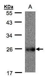
Sample(30 ug whole cell lysate) A: Raji(GTX27908) 12% SDS PAGE GTX102150 diluted at 1:500
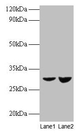
 A: Mouse brain 12% SDS PAGE GTX102150 diluted at 1:1000")
![UQCRFS1 antibody [N1C3] detects UQCRFS1 protein at mitochondria by immunofluorescent analysis. Sample: A549 cells were fixed in 2% paraformaldehyde/culture medium at 37oC for 30 min. Green: UQCRFS1 protein stained by UQCRFS1 antibody [N1C3] (GTX102150) diluted at 1:500. Blue: Hoechst 33342 staining. Scale bar = 10 μm.](https://www.genetex.com/upload/website/prouct_img/normal/GTX102150/GTX102150_39918_IFA_w_23060100_239.webp "UQCRFS1 antibody [N1C3] detects UQCRFS1 protein at mitochondria by immunofluorescent analysis. Sample: A549 cells were fixed in 2% paraformaldehyde/culture medium at 37oC for 30 min. Green: UQCRFS1 protein stained by UQCRFS1 antibody [N1C3] (GTX102150) diluted at 1:500. Blue: Hoechst 33342 staining. Scale bar = 10 μm.")
![UQCRFS1 antibody [N1C3] detects UQCRFS1 protein at mitochondria by immunofluorescent analysis. Sample: H1299 cells were fixed in 2% paraformaldehyde/culture medium at 37oC for 30 min. Green: UQCRFS1 protein stained by UQCRFS1 antibody [N1C3] (GTX102150) diluted at 1:500. Blue: Hoechst 33342 staining.](https://www.genetex.com/upload/website/prouct_img/normal/GTX102150/GTX102150_39918_IFA_2_w_23060100_842.webp "UQCRFS1 antibody [N1C3] detects UQCRFS1 protein at mitochondria by immunofluorescent analysis. Sample: H1299 cells were fixed in 2% paraformaldehyde/culture medium at 37oC for 30 min. Green: UQCRFS1 protein stained by UQCRFS1 antibody [N1C3] (GTX102150) diluted at 1:500. Blue: Hoechst 33342 staining.")
Sample(30 ug whole cell lysate) A: Raji(GTX27908) 12% SDS PAGE GTX102150 diluted at 1:500
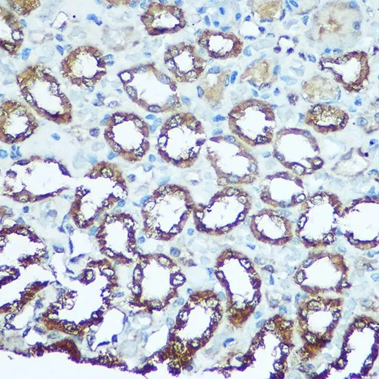
UQCRFS1 antibody [N1C3]
GTX102150
ApplicationsImmunoFluorescence, Western Blot, ImmunoCytoChemistry
Product group Antibodies
ReactivityHuman, Mouse
TargetUQCRFS1
Overview
- SupplierGeneTex
- Product NameUQCRFS1 antibody [N1C3]
- Delivery Days Customer9
- Application Supplier NoteWB: 1:500-1:3000. ICC/IF: 1:100-1:1000. *Optimal dilutions/concentrations should be determined by the researcher.Not tested in other applications.
- ApplicationsImmunoFluorescence, Western Blot, ImmunoCytoChemistry
- CertificationResearch Use Only
- ClonalityPolyclonal
- Concentration0.32 mg/ml
- ConjugateUnconjugated
- Gene ID7386
- Target nameUQCRFS1
- Target descriptionubiquinol-cytochrome c reductase, Rieske iron-sulfur polypeptide 1
- Target synonymsMC3DN10, RIP1, RIS1, RISP, UQCR5, cytochrome b-c1 complex subunit Rieske, mitochondrial, Rieske iron-sulfur protein, Rieske protein UQCRFS1, complex III subunit 5, cytochrome b-c1 complex subunit 5, epididymis secretory sperm binding protein, ubiquinol-cytochrome c reductase iron-sulfur subunit
- HostRabbit
- IsotypeIgG
- Protein IDP47985
- Protein NameCytochrome b-c1 complex subunit Rieske, mitochondrial
- Scientific DescriptionComponent of the ubiquinol-cytochrome c reductase complex (complex III or cytochrome b-c1 complex), which is a respiratory chain that generates an electrochemical potential coupled to ATP synthesis. The transit peptide of the Rieske protein seems to form part of the bc1 complex and is considered to be the subunit 11/IX of that complex.
- ReactivityHuman, Mouse
- Storage Instruction-20°C or -80°C,2°C to 8°C
- UNSPSC41116161
Datasheet
Related products
Product group Antibodies
Anti-UQCRFS1 AntibodyA31603
ApplicationsWestern Blot, ImmunoHistoChemistry
ReactivityHuman, Mouse, Rat
- SizePrice
Product group Antibodies

UQCRFS1 AntibodyLS-C749084
ApplicationsWestern Blot, ImmunoHistoChemistry
ReactivityHuman, Mouse
TargetUQCRFS1
- SizePrice

Product group Antibodies
ApplicationsFlow Cytometry, ImmunoFluorescence, ImmunoPrecipitation, Western Blot, ImmunoCytoChemistry, ImmunoHistoChemistry
ReactivityHuman, Mouse, Rat
TargetUQCRFS1
- SizePrice
Product group Antibodies
TargetUQCRFS1
- SizePrice
Product group Antibodies
UQCRFS1 AntibodyCSB-PA04645A0RB
ApplicationsWestern Blot, ELISA, ImmunoHistoChemistry
ReactivityHuman
TargetUQCRFS1
- SizePrice

Product group Antibodies
Anti-UQCRFS1 AntibodyHPA041863
ApplicationsWestern Blot, ImmunoCytoChemistry, ImmunoHistoChemistry
ReactivityHuman, Rat
TargetUQCRFS1
- SizePrice
Product group Antibodies
UQCRFS1 Polyclonal AntibodyCAC14065
ApplicationsWestern Blot, ELISA, ImmunoHistoChemistry
TargetUQCRFS1
- SizePrice
Product group Antibodies
UQCRFS1 Recombinant AntibodyBSM-54363R
ApplicationsFlow Cytometry, ImmunoFluorescence, Western Blot, ImmunoCytoChemistry, ImmunoHistoChemistry, ImmunoHistoChemistry Paraffin
ReactivityHuman, Mouse, Rat
TargetUQCRFS1
- SizePrice
Product group Antibodies
UQCRFS1 antibodyGTX33574
ApplicationsImmunoFluorescence, Western Blot, ImmunoCytoChemistry, ImmunoHistoChemistry, ImmunoHistoChemistry Paraffin
ReactivityHuman, Mouse, Rat
TargetUQCRFS1
- SizePrice