


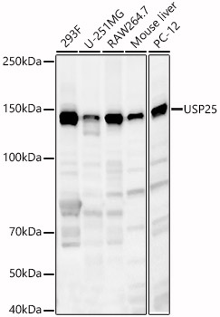
Figure 1. Western blot analysis of USP25 using anti-USP25 antibody (A06182-1). Electrophoresis was performed on a 5-20% SDS-PAGE gel at 70V (Stacking gel) / 90V (Resolving gel) for 2-3 hours. The sample well of each lane was loaded with 50ug of sample under reducing conditions. Lane 1: human HEK293 whole cell lysates, Lane 2: human placenta tissue lysates, Lane 3: monkey liver tissue lysates, Lane 4: monkey kidney tissue lysates, Lane 5: human PC-3 whole cell lysates, Lane 6: human Hela whole cell lysates, Lane 7: human A549 whole cell lysates, Lane 8: rat kidney tissue lysates, Lane 9: rat liver tissue lysates, Lane 10: rat lung tissue lysates, Lane 11: human PC-12 whole cell lysates, Lane 12: mouse lung tissue lysates, Lane 13: mouse RAW264.7 whole cell lysates, Lane 14: mouse NIH-3T3 whole cell lysates, After Electrophoresis, proteins were transferred to a Nitrocellulose membrane at 150mA for 50-90 minutes. Blocked the membrane with 5% Non-fat Milk/ TBS for 1.5 hour at RT. The membrane was incubated with rabbit anti-USP25 antigen affinity purified polyclonal antibody (Catalog # A06182-1) at 0.25 microg/mL overnight at 4°C, then washed with TBS-0.1%Tween 3 times with 5 minutes each and probed with a goat anti-rabbit IgG-HRP secondary antibody at a dilution of 1:5000 for 1.5 hour at RT. The signal is developed using an Enhanced Chemiluminescent detection (ECL) kit (Catalog # EK1002) with Tanon 5200 system. A specific band was detected for USP25 at approximately 125KD. The expected band size for USP25 is at 125KD.
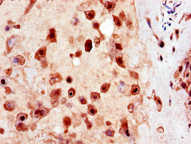
. USP25 was detected in immunocytochemical section of A549 cells. Enzyme antigen retrieval was performed using IHC enzyme antigen retrieval reagent (AR0022) for 15 mins. The cells were blocked with 10% goat serum. And then incubated with 2microg/mL rabbit anti-USP25 Antibody (A06182-1) overnight at 4°C. DyLight®488 Conjugated Goat Anti-Rabbit IgG (BA1127) was used as secondary antibody at 1:100 dilution and incubated for 30 minutes at 37°C. The section was counterstained with DAPI. Visualize using a fluorescence microscope and filter sets appropriate for the label used.")
. Overlay histogram showing HL-60 cells stained with A06182-1 (Blue line). To facilitate intracellular staining, cells were fixed with 4% paraformaldehyde and permeabilized with permeabilization buffer. The cells were blocked with 10% normal goat serum. And then incubated with rabbit anti-USP25 Antibody (A06182-1, 1microg/1x106 cells) for 30 min at 20°C. DyLight®488 conjugated goat anti-rabbit IgG (BA1127, 5-10microg/1x106 cells) was used as secondary antibody for 30 minutes at 20°C. Isotype control antibody (Green line) was rabbit IgG (1microg/1x106) used under the same conditions. Unlabelled sample without incubation with primary antibody and secondary antibody (Red line) was used as a blank control.")
Figure 1. Western blot analysis of USP25 using anti-USP25 antibody (A06182-1). Electrophoresis was performed on a 5-20% SDS-PAGE gel at 70V (Stacking gel) / 90V (Resolving gel) for 2-3 hours. The sample well of each lane was loaded with 50ug of sample under reducing conditions. Lane 1: human HEK293 whole cell lysates, Lane 2: human placenta tissue lysates, Lane 3: monkey liver tissue lysates, Lane 4: monkey kidney tissue lysates, Lane 5: human PC-3 whole cell lysates, Lane 6: human Hela whole cell lysates, Lane 7: human A549 whole cell lysates, Lane 8: rat kidney tissue lysates, Lane 9: rat liver tissue lysates, Lane 10: rat lung tissue lysates, Lane 11: human PC-12 whole cell lysates, Lane 12: mouse lung tissue lysates, Lane 13: mouse RAW264.7 whole cell lysates, Lane 14: mouse NIH-3T3 whole cell lysates, After Electrophoresis, proteins were transferred to a Nitrocellulose membrane at 150mA for 50-90 minutes. Blocked the membrane with 5% Non-fat Milk/ TBS for 1.5 hour at RT. The membrane was incubated with rabbit anti-USP25 antigen affinity purified polyclonal antibody (Catalog # A06182-1) at 0.25 microg/mL overnight at 4°C, then washed with TBS-0.1%Tween 3 times with 5 minutes each and probed with a goat anti-rabbit IgG-HRP secondary antibody at a dilution of 1:5000 for 1.5 hour at RT. The signal is developed using an Enhanced Chemiluminescent detection (ECL) kit (Catalog # EK1002) with Tanon 5200 system. A specific band was detected for USP25 at approximately 125KD. The expected band size for USP25 is at 125KD.
Anti-USP25 Antibody Picoband(r)
A06182-1-CARRIER-FREE
ApplicationsFlow Cytometry, ImmunoFluorescence, Western Blot, ELISA, ImmunoCytoChemistry
Product group Antibodies
ReactivityHuman, Monkey, Mouse, Rat
TargetUSP25
Overview
- SupplierBoster Bio
- Product NameAnti-USP25 Antibody Picoband(r)
- Delivery Days Customer9
- ApplicationsFlow Cytometry, ImmunoFluorescence, Western Blot, ELISA, ImmunoCytoChemistry
- CertificationResearch Use Only
- ClonalityPolyclonal
- Concentration500 ug/ml
- Gene ID29761
- Target nameUSP25
- Target descriptionubiquitin specific peptidase 25
- Target synonymsEIG19, USP21, ubiquitin carboxyl-terminal hydrolase 25, USP on chromosome 21, deubiquitinating enzyme 25, ubiquitin thioesterase 25, ubiquitin thiolesterase 25, ubiquitin-specific processing protease 25
- HostRabbit
- IsotypeIgG
- Protein IDQ9UHP3
- Protein NameUbiquitin carboxyl-terminal hydrolase 25
- Scientific DescriptionBoster Bio Anti-USP25 Antibody Picoband® catalog # A06182-1. Tested in ELISA, Flow Cytometry, IF, ICC, WB applications. This antibody reacts with Human, Monkey, Mouse, Rat. The brand Picoband indicates this is a premium antibody that guarantees superior quality, high affinity, and strong signals with minimal background in Western blot applications. Only our best-performing antibodies are designated as Picoband, ensuring unmatched performance.
- ReactivityHuman, Monkey, Mouse, Rat
- Storage Instruction-20°C,2°C to 8°C
- UNSPSC12352203
Related products
Product group Antibodies
Anti-USP25 AntibodyA88148
ApplicationsWestern Blot, ImmunoHistoChemistry
ReactivityHuman, Mouse, Rat
- SizePrice
Product group Antibodies

USP25 AntibodyLS-C830405
ApplicationsELISA, ImmunoHistoChemistry
ReactivityHuman, Mouse
TargetUSP25
- SizePrice

Product group Antibodies
Anti-USP25 AntibodyHPA018297
ApplicationsWestern Blot, ImmunoHistoChemistry
ReactivityHuman
TargetUSP25
- SizePrice
Product group Antibodies
USP25 AntibodyCSB-PA883400LA01HU
ApplicationsImmunoFluorescence, ELISA, ImmunoHistoChemistry
ReactivityHuman
TargetUSP25
- SizePrice
Product group Antibodies
Anti-USP25Y158094
ApplicationsWestern Blot, ELISA, ImmunoHistoChemistry
ReactivityHuman, Mouse, Rat
- SizePrice
Product group Antibodies
USP25 antibodyGTX17216
ApplicationsWestern Blot, ELISA, ImmunoHistoChemistry, ImmunoHistoChemistry Paraffin
ReactivityHuman, Mouse
TargetUSP25
- SizePrice
Product group Antibodies
Anti-USP25 Antibody144-12588
ApplicationsWestern Blot, ImmunoHistoChemistry
ReactivityHuman, Mouse, Rat
TargetUSP25
- SizePrice
Product group Antibodies
USP25 (7C1) Monoclonal AntibodyBSM-51259M
ApplicationsWestern Blot
ReactivityHuman
TargetUSP25
- SizePrice