

Anti-VDAC1 Antibody
144-00810
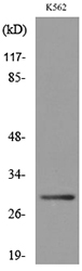
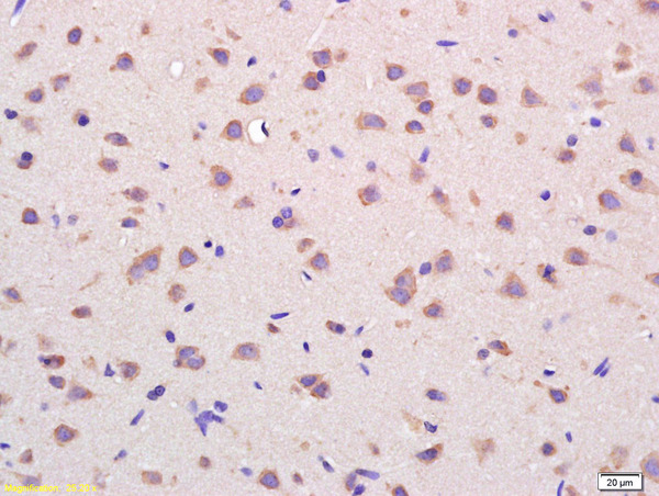
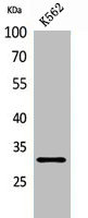
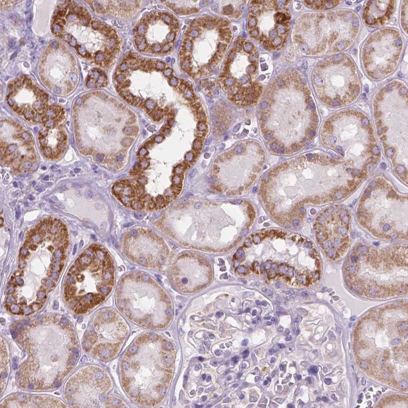
ApplicationsWestern Blot, ImmunoHistoChemistry
Product group Antibodies
ReactivityHuman, Mouse, Rat
TargetVDAC1
Overview
- SupplierRayBiotech
- Product NameAnti-VDAC1 Antibody
- Delivery Days Customer16
- ApplicationsWestern Blot, ImmunoHistoChemistry
- CertificationResearch Use Only
- ClonalityPolyclonal
- ConjugateUnconjugated
- Gene ID7416
- Target nameVDAC1
- Target descriptionvoltage dependent anion channel 1
- Target synonymsPORIN, VDAC-1, non-selective voltage-gated ion channel VDAC1, outer mitochondrial membrane protein porin 1, plasmalemmal porin, porin 31HL, porin 31HM, sperm binding protein 1a
- HostRabbit
- IsotypeIgG
- Protein IDP21796
- Protein NameNon-selective voltage-gated ion channel VDAC1
- Scientific DescriptionVDAC1 Polyclonal Antibody
- ReactivityHuman, Mouse, Rat
- Storage Instruction-20°C
- UNSPSC12352203
Related products
Product group Antibodies
Anti-VDAC1 AntibodyA97114
ApplicationsWestern Blot, ELISA
ReactivityHuman, Mouse, Rat
- SizePrice

Product group Antibodies
Anti-Porin/VDAC1 Antibody Picoband(r)A01168-1-CARRIER-FREE
ApplicationsFlow Cytometry, ImmunoFluorescence, Western Blot, ELISA, ImmunoCytoChemistry, ImmunoHistoChemistry
ReactivityHuman, Mouse, Rat
TargetVDAC1
- SizePrice
Product group Antibodies

VDAC1 / PORIN AntibodyLS-C831437
ApplicationsImmunoHistoChemistry
ReactivityHuman, Mouse, Rat
TargetVDAC1
- SizePrice
Product group Antibodies
References
VDAC Polyclonal AntibodyBS-1461R
ApplicationsImmunoFluorescence, Western Blot, ELISA, ImmunoCytoChemistry, ImmunoHistoChemistry, ImmunoHistoChemistry Frozen, ImmunoHistoChemistry Paraffin
ReactivityBovine, Canine, Equine, Human, Mouse, Porcine, Rabbit, Rat, Sheep
TargetVDAC1
- SizePrice
Product group Antibodies
Vdac1 Polyclonal AntibodyCAC11640
ApplicationsWestern Blot, ELISA
ReactivityPlant
- SizePrice
Product group Antibodies
VDAC1 AntibodyCSB-PA005626
ApplicationsWestern Blot, ELISA
ReactivityHuman, Mouse, Rat
TargetVDAC1
- SizePrice
Product group Antibodies
Anti-VDAC1 AntibodyHPA030780
ApplicationsWestern Blot, ImmunoHistoChemistry
ReactivityHuman
TargetVDAC1
- SizePrice
![VDAC1 antibody [N1C2] detects VDAC1 protein at mitochondria on mouse muscle by immunohistochemical analysis. Sample: Paraffin-embedded mouse muscle. VDAC1 antibody [N1C2] (GTX114187) dilution: 1:500.
Antigen Retrieval: Trilogy? (EDTA based, pH 8.0) buffer, 15min](https://www.genetex.com/upload/website/prouct_img/normal/GTX114187/GTX114187_40156_IHC_M_w_23060501_567.webp)
Product group Antibodies
VDAC1 antibody [N1C2]GTX114187
ApplicationsImmunoFluorescence, Western Blot, ImmunoCytoChemistry, ImmunoHistoChemistry, ImmunoHistoChemistry Paraffin
ReactivityHuman, Mouse, Rat
TargetVDAC1
- SizePrice