

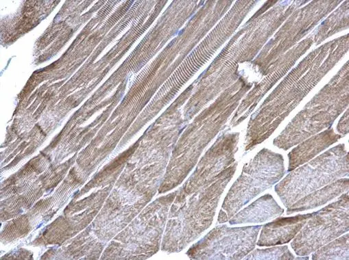
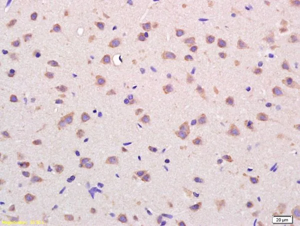
VDAC1 antibody [N1C2] detects VDAC1 protein at mitochondria on mouse muscle by immunohistochemical analysis. Sample: Paraffin-embedded mouse muscle. VDAC1 antibody [N1C2] (GTX114187) dilution: 1:500.
Antigen Retrieval: Trilogy? (EDTA based, pH 8.0) buffer, 15min
 antibody at 1:500 dilution.
Antigen Retrieval: Trilogy? (EDTA based, pH 8.0) buffer, 15min")

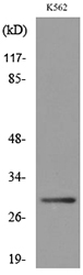
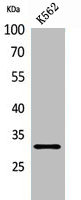
![Various whole cell extracts (30 μg) were separated by 12% SDS-PAGE, and the membrane was blotted with VDAC1 antibody [N1C2] (GTX114187) diluted at 1:1000. The HRP-conjugated anti-rabbit IgG antibody (GTX213110-01) was used to detect the primary antibody.](https://www.genetex.com/upload/website/prouct_img/normal/GTX114187/GTX114187_45462_20240729_WB_24082301_494.webp "Various whole cell extracts (30 μg) were separated by 12% SDS-PAGE, and the membrane was blotted with VDAC1 antibody [N1C2] (GTX114187) diluted at 1:1000. The HRP-conjugated anti-rabbit IgG antibody (GTX213110-01) was used to detect the primary antibody.")
VDAC1 antibody [N1C2] detects VDAC1 protein at mitochondria on mouse muscle by immunohistochemical analysis. Sample: Paraffin-embedded mouse muscle. VDAC1 antibody [N1C2] (GTX114187) dilution: 1:500.
Antigen Retrieval: Trilogy? (EDTA based, pH 8.0) buffer, 15min
VDAC1 antibody [N1C2]
GTX114187
ApplicationsImmunoFluorescence, Western Blot, ImmunoCytoChemistry, ImmunoHistoChemistry, ImmunoHistoChemistry Paraffin
Product group Antibodies
ReactivityHuman, Mouse, Rat
TargetVDAC1
Overview
- SupplierGeneTex
- Product NameVDAC1 antibody [N1C2]
- Delivery Days Customer9
- Application Supplier NoteWB: 1:500-1:3000. IHC-P: 1:100-1:1000. *Optimal dilutions/concentrations should be determined by the researcher.Not tested in other applications.
- ApplicationsImmunoFluorescence, Western Blot, ImmunoCytoChemistry, ImmunoHistoChemistry, ImmunoHistoChemistry Paraffin
- CertificationResearch Use Only
- ClonalityPolyclonal
- Concentration1.46 mg/ml
- ConjugateUnconjugated
- Gene ID7416
- Target nameVDAC1
- Target descriptionvoltage dependent anion channel 1
- Target synonymsPORIN, VDAC-1, non-selective voltage-gated ion channel VDAC1, outer mitochondrial membrane protein porin 1, plasmalemmal porin, porin 31HL, porin 31HM, sperm binding protein 1a
- HostRabbit
- IsotypeIgG
- Protein IDP21796
- Protein NameNon-selective voltage-gated ion channel VDAC1
- Scientific DescriptionThis gene encodes a voltage-dependent anion channel protein that is a major component of the outer mitochondrial membrane. The encoded protein facilitates the exchange of metabolites and ions across the outer mitochondrial membrane and may regulate mitochondrial functions. This protein also forms channels in the plasma membrane and may be involved in transmembrane electron transport. Alternate splicing results in multiple transcript variants. Multiple pseudogenes of this gene are found on chromosomes 1, 2 3, 6, 9, 12, X and Y.
- ReactivityHuman, Mouse, Rat
- Storage Instruction-20°C or -80°C,2°C to 8°C
- UNSPSC41116161
Datasheet
Related products
Product group Antibodies
Anti-VDAC1 AntibodyA97114
ApplicationsWestern Blot, ELISA
ReactivityHuman, Mouse, Rat
- SizePrice

Product group Antibodies
Anti-Porin/VDAC1 Antibody Picoband(r)A01168-1-CARRIER-FREE
ApplicationsFlow Cytometry, ImmunoFluorescence, Western Blot, ELISA, ImmunoCytoChemistry, ImmunoHistoChemistry
ReactivityHuman, Mouse, Rat
TargetVDAC1
- SizePrice
Product group Antibodies
Anti-VDAC1 Antibody144-00810
ApplicationsWestern Blot, ImmunoHistoChemistry
ReactivityHuman, Mouse, Rat
TargetVDAC1
- SizePrice
Product group Antibodies

VDAC1 / PORIN AntibodyLS-C831437
ApplicationsImmunoHistoChemistry
ReactivityHuman, Mouse, Rat
TargetVDAC1
- SizePrice
Product group Antibodies
References
VDAC Polyclonal AntibodyBS-1461R
ApplicationsImmunoFluorescence, Western Blot, ELISA, ImmunoCytoChemistry, ImmunoHistoChemistry, ImmunoHistoChemistry Frozen, ImmunoHistoChemistry Paraffin
ReactivityBovine, Canine, Equine, Human, Mouse, Porcine, Rabbit, Rat, Sheep
TargetVDAC1
- SizePrice
Product group Antibodies
Vdac1 Polyclonal AntibodyCAC11640
ApplicationsWestern Blot, ELISA
ReactivityPlant
- SizePrice
Product group Antibodies
VDAC1 AntibodyCSB-PA005626
ApplicationsWestern Blot, ELISA
ReactivityHuman, Mouse, Rat
TargetVDAC1
- SizePrice
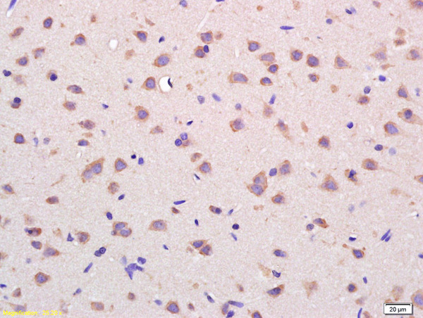
![IHC-P analysis of human colon carcinoma tissue section using GTX01543 VDAC1 antibody [GT1235]. Dilution : 1:100](https://www.genetex.com/upload/website/prouct_img/normal/GTX01543/GTX01543_20200508_IHC-P_w_23053121_813.webp)
Product group Antibodies
VDAC1 antibody [GT1235]GTX01543
ApplicationsImmunoFluorescence, Western Blot, ImmunoCytoChemistry, ImmunoHistoChemistry, ImmunoHistoChemistry Paraffin
ReactivityHuman, Mouse, Rat
TargetVDAC1
- SizePrice
Product group Antibodies
VDAC1 antibodyGTX03668
ApplicationsImmunoPrecipitation, Western Blot, ImmunoHistoChemistry, ImmunoHistoChemistry Paraffin
ReactivityHuman, Mouse, Rat
TargetVDAC1
- SizePrice
Product group Antibodies
Anti-VDAC1 AntibodyHPA030780
ApplicationsWestern Blot, ImmunoHistoChemistry
ReactivityHuman
TargetVDAC1
- SizePrice