

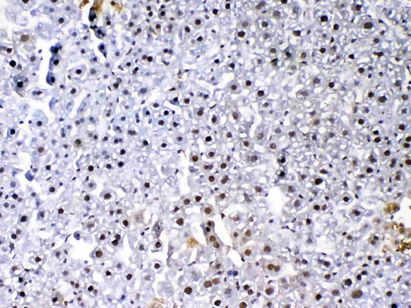
Figure 7. IHC analysis of VEGF Receptor 3 using anti-VEGF Receptor 3 antibody (A01276-3). VEGF Receptor 3 was detected in paraffin-embedded section of mouse liver tissue. Heat mediated antigen retrieval was performed in citrate buffer (pH6, epitope retrieval solution) for 20 mins. The tissue section was blocked with 10% goat serum. The tissue section was then incubated with 1microg/ml rabbit anti-VEGF Receptor 3 Antibody (A01276-3) overnight at 4°C. Biotinylated goat anti-rabbit IgG was used as secondary antibody and incubated for 30 minutes at 37°C. The tissue section was developed using Strepavidin-Biotin-Complex (SABC)(Catalog # SA1022) with DAB as the chromogen.
. VEGF Receptor 3 was detected in paraffin-embedded section of rat liver tissue. Heat mediated antigen retrieval was performed in citrate buffer (pH6, epitope retrieval solution) for 20 mins. The tissue section was blocked with 10% goat serum. The tissue section was then incubated with 1microg/ml rabbit anti-VEGF Receptor 3 Antibody (A01276-3) overnight at 4°C. Biotinylated goat anti-rabbit IgG was used as secondary antibody and incubated for 30 minutes at 37°C. The tissue section was developed using Strepavidin-Biotin-Complex (SABC)(Catalog # SA1022) with DAB as the chromogen.")
. VEGF Receptor 3 was detected in paraffin-embedded section of human rectal cancer tissue. Heat mediated antigen retrieval was performed in citrate buffer (pH6, epitope retrieval solution) for 20 mins. The tissue section was blocked with 10% goat serum. The tissue section was then incubated with 1microg/ml rabbit anti-VEGF Receptor 3 Antibody (A01276-3) overnight at 4°C. Biotinylated goat anti-rabbit IgG was used as secondary antibody and incubated for 30 minutes at 37°C. The tissue section was developed using Strepavidin-Biotin-Complex (SABC)(Catalog # SA1022) with DAB as the chromogen.")
. VEGF Receptor 3 was detected in paraffin-embedded section of human prostatic cancer tissue. Heat mediated antigen retrieval was performed in citrate buffer (pH6, epitope retrieval solution) for 20 mins. The tissue section was blocked with 10% goat serum. The tissue section was then incubated with 1microg/ml rabbit anti-VEGF Receptor 3 Antibody (A01276-3) overnight at 4°C. Biotinylated goat anti-rabbit IgG was used as secondary antibody and incubated for 30 minutes at 37°C. The tissue section was developed using Strepavidin-Biotin-Complex (SABC)(Catalog # SA1022) with DAB as the chromogen.")
. VEGF Receptor 3 was detected in paraffin-embedded section of human lung cancer tissue. Heat mediated antigen retrieval was performed in citrate buffer (pH6, epitope retrieval solution) for 20 mins. The tissue section was blocked with 10% goat serum. The tissue section was then incubated with 1microg/ml rabbit anti-VEGF Receptor 3 Antibody (A01276-3) overnight at 4°C. Biotinylated goat anti-rabbit IgG was used as secondary antibody and incubated for 30 minutes at 37°C. The tissue section was developed using Strepavidin-Biotin-Complex (SABC)(Catalog # SA1022) with DAB as the chromogen.")
. VEGF Receptor 3 was detected in paraffin-embedded section of human placenta tissue. Heat mediated antigen retrieval was performed in citrate buffer (pH6, epitope retrieval solution) for 20 mins. The tissue section was blocked with 10% goat serum. The tissue section was then incubated with 1microg/ml rabbit anti-VEGF Receptor 3 Antibody (A01276-3) overnight at 4°C. Biotinylated goat anti-rabbit IgG was used as secondary antibody and incubated for 30 minutes at 37°C. The tissue section was developed using Strepavidin-Biotin-Complex (SABC)(Catalog # SA1022) with DAB as the chromogen.")
. Electrophoresis was performed on a 5-20% SDS-PAGE gel at 70V (Stacking gel) / 90V (Resolving gel) for 2-3 hours. The sample well of each lane was loaded with 50ug of sample under reducing conditions. Lane 1: rat liver tissue lysates. After Electrophoresis, proteins were transferred to a Nitrocellulose membrane at 150mA for 50-90 minutes. Blocked the membrane with 5% Non-fat Milk/ TBS for 1.5 hour at RT. The membrane was incubated with rabbit anti-VEGF Receptor 3 antigen affinity purified polyclonal antibody (Catalog # A01276-3) at 0.5 microg/mL overnight at 4°C, then washed with TBS-0.1%Tween 3 times with 5 minutes each and probed with a goat anti-rabbit IgG-HRP secondary antibody at a dilution of 1:10000 for 1.5 hour at RT. The signal is developed using an Enhanced Chemiluminescent detection (ECL) kit (Catalog # EK1002) with Tanon 5200 system. A specific band was detected for VEGF Receptor 3 at approximately 153KD. The expected band size for VEGF Receptor 3 is at 153KD.")
. VEGF Receptor 3 was detected in frozen section of human placenta tissue. The tissue section was blocked with 10% goat serum. The tissue section was then incubated with 1microg/ml rabbit anti-VEGF Receptor 3 Antibody (A01276-3) overnight at 4°C. Biotinylated goat anti-rabbit IgG was used as secondary antibody and incubated for 30 minutes at 37°C. The tissue section was developed using Strepavidin-Biotin-Complex (SABC)(Catalog # SA1022) with DAB as the chromogen.")
. VEGF Receptor 3 was detected in immunocytochemical section of A431 cells. Enzyme antigen retrieval was performed using IHC enzyme antigen retrieval reagent (AR0022) for 15 mins. The cells were blocked with 10% goat serum. And then incubated with 2microg/mL rabbit anti-VEGF Receptor 3 Antibody (A01276-3) overnight at 4°C. DyLight®488 Conjugated Goat Anti-Rabbit IgG (BA1127) was used as secondary antibody at 1:100 dilution and incubated for 30 minutes at 37°C. The section was counterstained with DAPI. Visualize using a fluorescence microscope and filter sets appropriate for the label used.")
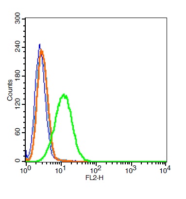
. Overlay histogram showing U20S cells stained with A01276-3 (Blue line). To facilitate intracellular staining, cells were fixed with 4% paraformaldehyde and permeabilized with permeabilization buffer. The cells were blocked with 10% normal goat serum. And then incubated with rabbit anti-VEGF Receptor 3 Antibody (A01276-3,1microg/1x106 cells) for 30 min at 20°C. DyLight®488 conjugated goat anti-rabbit IgG (BA1127, 5-10microg/1x106 cells) was used as secondary antibody for 30 minutes at 20°C. Isotype control antibody (Green line) was rabbit IgG (1microg/1x106) used under the same conditions. Unlabelled sample without incubation with primary antibody and secondary antibody (Red line) was used as a blank control.")
Figure 7. IHC analysis of VEGF Receptor 3 using anti-VEGF Receptor 3 antibody (A01276-3). VEGF Receptor 3 was detected in paraffin-embedded section of mouse liver tissue. Heat mediated antigen retrieval was performed in citrate buffer (pH6, epitope retrieval solution) for 20 mins. The tissue section was blocked with 10% goat serum. The tissue section was then incubated with 1microg/ml rabbit anti-VEGF Receptor 3 Antibody (A01276-3) overnight at 4°C. Biotinylated goat anti-rabbit IgG was used as secondary antibody and incubated for 30 minutes at 37°C. The tissue section was developed using Strepavidin-Biotin-Complex (SABC)(Catalog # SA1022) with DAB as the chromogen.
Anti-VEGF Receptor 3/FLT4 Antibody Picoband(r)
A01276-3-PE
ApplicationsFlow Cytometry, ImmunoFluorescence, Western Blot, ELISA, ImmunoCytoChemistry, ImmunoHistoChemistry, ImmunoHistoChemistry Frozen
Product group Antibodies
ReactivityHuman, Mouse, Rat
TargetFLT4
Overview
- SupplierBoster Bio
- Product NameAnti-VEGF Receptor 3/FLT4 Antibody Picoband(r)
- Delivery Days Customer9
- ApplicationsFlow Cytometry, ImmunoFluorescence, Western Blot, ELISA, ImmunoCytoChemistry, ImmunoHistoChemistry, ImmunoHistoChemistry Frozen
- CertificationResearch Use Only
- ClonalityPolyclonal
- Concentration500 ug/ml
- ConjugateRPE
- Gene ID2324
- Target nameFLT4
- Target descriptionfms related receptor tyrosine kinase 4
- Target synonymsCHTD7, FLT-4, FLT41, LMPH1A, LMPHM1, PCL, VEGFR-3, VEGFR3, vascular endothelial growth factor receptor 3, Feline McDonough Sarcoma (FMS)-like tyrosine kinase 4, VEGF receptor-3, fms related tyrosine kinase 4, fms-like tyrosine kinase 4, primary congenital lymphedema, tyrosine-protein kinase receptor FLT4
- HostRabbit
- IsotypeIgG
- Protein IDP35916
- Protein NameVascular endothelial growth factor receptor 3
- Scientific DescriptionBoster Bio Anti-VEGF Receptor 3/FLT4 Antibody Picoband® catalog # A01276-3. Tested in ELISA, Flow Cytometry, IF, IHC, IHC-F, ICC, WB applications. This antibody reacts with Human, Mouse, Rat. The brand Picoband indicates this is a premium antibody that guarantees superior quality, high affinity, and strong signals with minimal background in Western blot applications. Only our best-performing antibodies are designated as Picoband, ensuring unmatched performance.
- ReactivityHuman, Mouse, Rat
- Storage Instruction-20°C,2°C to 8°C
- UNSPSC12352203
Related products

Product group Antibodies
Anti-VEGF Receptor 3/FLT4 Antibody Picoband(r)A01276-2-CARRIER-FREE
ApplicationsFlow Cytometry, ImmunoFluorescence, Western Blot, ImmunoCytoChemistry, ImmunoHistoChemistry, ImmunoHistoChemistry Frozen
ReactivityHuman, Mouse, Rat
TargetFLT4
- SizePrice
Product group Antibodies

Anti-VEGFR-3 [ABDD073]AB04028-10.0
ApplicationsFlow Cytometry, ELISA, ImmunoHistoChemistry
ReactivityHuman
TargetFLT4
- SizePrice
Product group Antibodies
Anti-VEGFR3 AntibodyA34796
ApplicationsImmunoFluorescence, Western Blot, ImmunoHistoChemistry
ReactivityHuman
- SizePrice
Product group Antibodies

FLT4 / VEGFR3 AntibodyLS-C832540
ApplicationsImmunoFluorescence, ELISA
ReactivityHuman
TargetFLT4
- SizePrice
Product group Antibodies
Phospho-KDR/FLT4 (Y1054/Y1063) AntibodyCSB-PA009643
ApplicationsELISA, ImmunoHistoChemistry
ReactivityHuman, Mouse, Rat
TargetFLT4
- SizePrice

Product group Antibodies
Anti-FLT4 AntibodyHPA046519
ApplicationsImmunoCytoChemistry
ReactivityHuman
TargetFLT4
- SizePrice
Product group Antibodies
ApplicationsImmunoPrecipitation, Western Blot, ImmunoCytoChemistry, ImmunoHistoChemistry
TargetFLT4
- SizePrice
Product group Antibodies
References
VEGFR3 Polyclonal AntibodyBS-2202R
ApplicationsFlow Cytometry, ImmunoFluorescence, ELISA, ImmunoCytoChemistry, ImmunoHistoChemistry, ImmunoHistoChemistry Frozen, ImmunoHistoChemistry Paraffin
ReactivityCanine, Equine, Human, Mouse, Porcine, Rabbit, Rat
TargetFLT4
- SizePrice
Product group Antibodies
VEGF Receptor 3 antibodyGTX100807
ApplicationsWestern Blot
ReactivityHuman, Mouse
TargetFLT4
- SizePrice
Product group Antibodies
Anti-Mouse/Rat FLT4 Antibody144-12332
ApplicationsWestern Blot
ReactivityHuman, Mouse, Rat
TargetFLT4
- SizePrice