


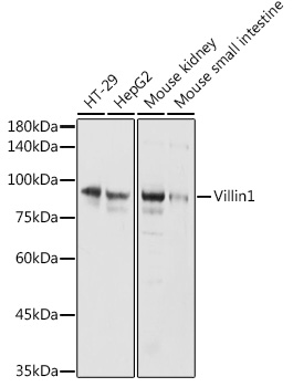
Figure 1. Western blot analysis of Villin using anti-Villin antibody (PB9457). Electrophoresis was performed on a 5-20% SDS-PAGE gel at 70V (Stacking gel) / 90V (Resolving gel) for 2-3 hours. Lane 1: Rat Intestine Tissue Lysate at 50ug Lane 2: Mouse Kidney Tissue Lysate at 50ug Lane 3: RH35 Whole Cell Lysate at 40ug Lane 4: HEPG2 Whole Cell Lysate at 40ug, Lane 5: MCF-7 Whole Cell Lysate at 40ug. After electrophoresis, proteins were transferred to a nitrocellulose membrane at 150 mA for 50-90 minutes. Blocked the membrane with 5% non-fat milk/TBS for 1.5 hour at RT. The membrane was incubated with rabbit anti-Villin antigen affinity purified polyclonal antibody (Catalog # PB9457) at 0.5 microg/mL overnight at 4°C, then washed with TBS-0.1%Tween 3 times with 5 minutes each and probed with a goat anti-rabbit IgG-HRP secondary antibody at a dilution of 1:5000 for 1.5 hour at RT. The signal is developed using an Enhanced Chemiluminescent detection (ECL) kit (Catalog # EK1002) with Tanon 5200 system. A specific band was detected for Villin at approximately 93 kDa. The expected band size for Villin is at 93 kDa.
. Villin was detected in a paraffin-embedded section of mouse intestine tissue. Heat mediated antigen retrieval was performed in EDTA buffer (pH 8.0, epitope retrieval solution). The tissue section was blocked with 10% goat serum. The tissue section was then incubated with 1 microg/ml rabbit anti-Villin Antibody (PB9457) overnight at 4°C. Biotinylated goat anti-rabbit IgG was used as secondary antibody and incubated for 30 minutes at 37°C. The tissue section was developed using Strepavidin-Biotin-Complex (SABC) (Catalog # SA1022) with DAB as the chromogen.")
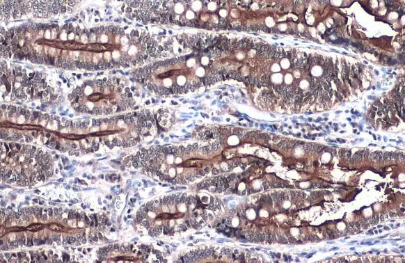
. Villin was detected in a paraffin-embedded section of rat intestine tissue. Heat mediated antigen retrieval was performed in EDTA buffer (pH 8.0, epitope retrieval solution). The tissue section was blocked with 10% goat serum. The tissue section was then incubated with 1 microg/ml rabbit anti-Villin Antibody (PB9457) overnight at 4°C. Biotinylated goat anti-rabbit IgG was used as secondary antibody and incubated for 30 minutes at 37°C. The tissue section was developed using Strepavidin-Biotin-Complex (SABC) (Catalog # SA1022) with DAB as the chromogen.")
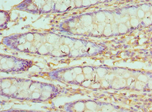
. Villin was detected in a paraffin-embedded section of human intestinal cancer tissue. Heat mediated antigen retrieval was performed in EDTA buffer (pH 8.0, epitope retrieval solution). The tissue section was blocked with 10% goat serum. The tissue section was then incubated with 1 microg/ml rabbit anti-Villin Antibody (PB9457) overnight at 4°C. Biotinylated goat anti-rabbit IgG was used as secondary antibody and incubated for 30 minutes at 37°C. The tissue section was developed using Strepavidin-Biotin-Complex (SABC) (Catalog # SA1022) with DAB as the chromogen.")
 Villi was detected in paraffin-embedded section of human rectal cancer tissues. Heat mediated antigen retrieval was performed in citrate buffer (pH6, epitope retrieval solution ) for 20 mins. The tissue section was blocked with 10% goat serum. The tissue section was then incubated with 1microg/mL rabbit anti-Villi Antibody (PB9457) overnight at 4°C. DyLight488 Conjugated Goat Anti-Rabbit IgG (BA1127) was used as secondary antibody at 1:100 dilution and incubated for 30 minutes at 37°C. The section was counterstained with DAPI. Visualize using a fluorescence microscope and filter sets appropriate for the label used.")
 Villi was detected in paraffin-embedded section of rat intestine tissues. Heat mediated antigen retrieval was performed in citrate buffer (pH6, epitope retrieval solution ) for 20 mins. The tissue section was blocked with 10% goat serum. The tissue section was then incubated with 1microg/mL rabbit anti-Villi Antibody (PB9457) overnight at 4°C. DyLight488 Conjugated Goat Anti-Rabbit IgG (BA1127) was used as secondary antibody at 1:100 dilution and incubated for 30 minutes at 37°C. The section was counterstained with DAPI. Visualize using a fluorescence microscope and filter sets appropriate for the label used.")
 Villi was detected in paraffin-embedded section of mouse intestine tissues. Heat mediated antigen retrieval was performed in citrate buffer (pH6, epitope retrieval solution ) for 20 mins. The tissue section was blocked with 10% goat serum. The tissue section was then incubated with 1microg/mL rabbit anti-Villi Antibody (PB9457) overnight at 4°C. DyLight488 Conjugated Goat Anti-Rabbit IgG (BA1127) was used as secondary antibody at 1:100 dilution and incubated for 30 minutes at 37°C. The section was counterstained with DAPI. Visualize using a fluorescence microscope and filter sets appropriate for the label used.")
. Villin was detected in paraffin-embedded section of human ileum tissue. Heat mediated antigen retrieval was performed in citrate buffer (pH6, epitope retrieval solution) for 20 mins. The tissue section was blocked with 10% goat serum. The tissue section was then incubated with 5microg/mL rabbit anti-Villin Antibody (PB9457) overnight at 4°C. DyLight®488 Conjugated Goat Anti-Rabbit IgG (BA1127) was used as secondary antibody at 1:100 dilution and incubated for 30 minutes at 37°C. The section was counterstained with DAPI. Visualize using a fluorescence microscope and filter sets appropriate for the label used.")
. Villin was detected in paraffin-embedded section of human colon organoid tissue. Heat mediated antigen retrieval was performed in citrate buffer (pH6, epitope retrieval solution) for 20 mins. The tissue section was blocked with 10% goat serum. The tissue section was then incubated with 5microg/mL rabbit anti-Villin Antibody (PB9457) overnight at 4°C. DyLight®488 Conjugated Goat Anti-Rabbit IgG (BA1127) was used as secondary antibody at 1:100 dilution and incubated for 30 minutes at 37°C. The section was counterstained with DAPI. Visualize using a fluorescence microscope and filter sets appropriate for the label used.")
. Villin was detected in paraffin-embedded section of mouse ileum tissue. Heat mediated antigen retrieval was performed in citrate buffer (pH6, epitope retrieval solution) for 20 mins. The tissue section was blocked with 10% goat serum. The tissue section was then incubated with 5microg/mL rabbit anti-Villin Antibody (PB9457) overnight at 4°C. DyLight®488 Conjugated Goat Anti-Rabbit IgG (BA1127) was used as secondary antibody at 1:100 dilution and incubated for 30 minutes at 37°C. The section was counterstained with DAPI. Visualize using a fluorescence microscope and filter sets appropriate for the label used.")
Figure 1. Western blot analysis of Villin using anti-Villin antibody (PB9457). Electrophoresis was performed on a 5-20% SDS-PAGE gel at 70V (Stacking gel) / 90V (Resolving gel) for 2-3 hours. Lane 1: Rat Intestine Tissue Lysate at 50ug Lane 2: Mouse Kidney Tissue Lysate at 50ug Lane 3: RH35 Whole Cell Lysate at 40ug Lane 4: HEPG2 Whole Cell Lysate at 40ug, Lane 5: MCF-7 Whole Cell Lysate at 40ug. After electrophoresis, proteins were transferred to a nitrocellulose membrane at 150 mA for 50-90 minutes. Blocked the membrane with 5% non-fat milk/TBS for 1.5 hour at RT. The membrane was incubated with rabbit anti-Villin antigen affinity purified polyclonal antibody (Catalog # PB9457) at 0.5 microg/mL overnight at 4°C, then washed with TBS-0.1%Tween 3 times with 5 minutes each and probed with a goat anti-rabbit IgG-HRP secondary antibody at a dilution of 1:5000 for 1.5 hour at RT. The signal is developed using an Enhanced Chemiluminescent detection (ECL) kit (Catalog # EK1002) with Tanon 5200 system. A specific band was detected for Villin at approximately 93 kDa. The expected band size for Villin is at 93 kDa.
Anti-Villin/VIL1 Antibody Picoband(r)
PB9457-CARRIER-FREE
ApplicationsFlow Cytometry, ImmunoFluorescence, Western Blot, ImmunoHistoChemistry
Product group Antibodies
ReactivityHamster, Human, Mouse, Rat
TargetVIL1
Overview
- SupplierBoster Bio
- Product NameAnti-Villin/VIL1 Antibody Picoband(r)
- Delivery Days Customer9
- Application Supplier NoteWB: The detection limit for Villin is approximately 0.1ng/lane under reducing conditions. Tested Species: In-house tested species with positive results. By Heat: Boiling the paraffin sections in 10mM citrate buffer, pH6.0, for 20mins is required for the staining of formalin/paraffin sections. Other applications have not been tested. Optimal dilutions should be determined by end users.
- ApplicationsFlow Cytometry, ImmunoFluorescence, Western Blot, ImmunoHistoChemistry
- CertificationResearch Use Only
- ClonalityPolyclonal
- Concentration500 ug/ml
- Gene ID7429
- Target nameVIL1
- Target descriptionvillin 1
- Target synonymsD2S1471, VIL, villin-1
- HostRabbit
- IsotypeIgG
- Protein IDP09327
- Protein NameVillin-1
- Scientific DescriptionBoster Bio Anti-Villin/VIL1 Antibody Picoband® catalog # PB9457. Tested in Flow Cytometry, IF, IHC, WB applications. This antibody reacts with Human, Mouse, Rat. The brand Picoband indicates this is a premium antibody that guarantees superior quality, high affinity, and strong signals with minimal background in Western blot applications. Only our best-performing antibodies are designated as Picoband, ensuring unmatched performance.
- ReactivityHamster, Human, Mouse, Rat
- Storage Instruction-20°C,2°C to 8°C
- UNSPSC12352203
Related products
Product group Antibodies
Anti-Villin AntibodyA14804
ApplicationsWestern Blot
ReactivityHuman, Mouse, Rat
- SizePrice

Product group Antibodies
villin Recombinant AntibodyBSM-54212R
ApplicationsFlow Cytometry, ImmunoFluorescence, Western Blot, ImmunoCytoChemistry, ImmunoHistoChemistry, ImmunoHistoChemistry Paraffin
ReactivityHuman, Mouse, Rat
TargetVIL1
- SizePrice
Product group Antibodies
Villin (VIL) Polyclonal AntibodyCAU24074
ApplicationsWestern Blot, ImmunoHistoChemistry
ReactivityMouse, Porcine, Rat
TargetVIL1
- SizePrice
Product group Antibodies
VIL1 AntibodyCSB-PA025855ESR1HU
ApplicationsELISA, ImmunoHistoChemistry
ReactivityHuman
TargetVIL1
- SizePrice

Product group Antibodies
Anti-VIL1 AntibodyHPA006884
ApplicationsWestern Blot, ImmunoCytoChemistry, ImmunoHistoChemistry
ReactivityHuman
TargetVIL1
- SizePrice
Product group Antibodies
Villin antibodyGTX109940
ApplicationsImmunoFluorescence, Western Blot, ImmunoCytoChemistry, ImmunoHistoChemistry, ImmunoHistoChemistry Frozen, ImmunoHistoChemistry Paraffin
ReactivityCanine, Feline, Human, Mouse, Rat
TargetVIL1
- SizePrice
Product group Antibodies
Anti-Villin1 AntibodyCAB5494
ApplicationsWestern Blot, ELISA
ReactivityHuman
TargetVIL1
- SizePrice
Product group Antibodies

VIL1 / Villin AntibodyLS-C783283
ApplicationsWestern Blot
ReactivityHuman
TargetVIL1
- SizePrice