

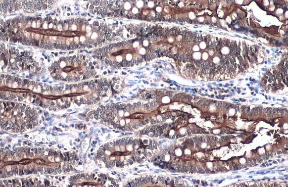
Villin antibody detects VIL1 protein at cell membrane by immunohistochemical analysis. Sample: Paraffin-embedded dog intestine. VIL1 stained by Villin antibody (GTX109940) diluted at 1:500. Antigen Retrieval: Citrate buffer, pH 6.0, 15 min
 diluted at 1:500. Antigen Retrieval: Citrate buffer, pH 6.0, 15 min")
 diluted at 1:500. Antigen Retrieval: Citrate buffer, pH 6.0, 15 min")
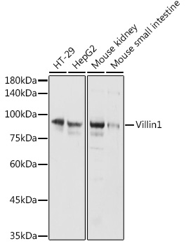
 were separated by 7.5% SDS-PAGE, and the membrane was blotted with Villin antibody (GTX109940) diluted at 1:500. The HRP-conjugated anti-rabbit IgG antibody (GTX213110-01) was used to detect the primary antibody.")
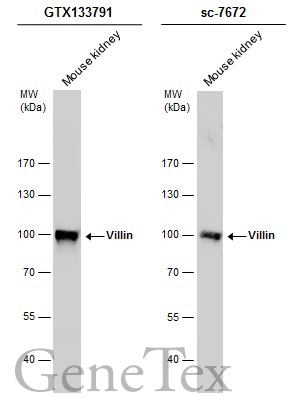
 was separated by 7.5% SDS-PAGE, and the membranes were blotted with Villin antibody (GTX109940) diluted at 1:5000 and competitor's antibody (sc-7672) diluted at 1:100. The HRP-conjugated anti-rabbit IgG antibody (GTX213110-01) was used to detect the primary antibody.")
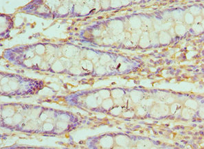
 antibody at 1:500 dilution.
Antigen Retrieval: Trilogy? (EDTA based, pH 8.0) buffer, 15min")
 diluted at 1:500. Antigen Retrieval: Citrate buffer, pH 6.0, 15 min")
 was separated by 7.5% SDS-PAGE, and the membrane was blotted with Villin antibody (GTX109940) diluted at 1:1000. The HRP-conjugated anti-rabbit IgG antibody (GTX213110-01) was used to detect the primary antibody.")
 dilution: 1:2000 The HRP-conjugated anti-rabbit IgG antibody (GTX213110-01) was used to detect the primary antibody.")
 were separated by 7.5% SDS-PAGE, and the membrane was blotted with Villin antibody (GTX109940) diluted at 1:2000. The HRP-conjugated anti-rabbit IgG antibody (GTX213110-01) was used to detect the primary antibody. Corresponding RNA expression data for the same cell lines are based on Human Protein Atlas program.")
Villin antibody detects VIL1 protein at cell membrane by immunohistochemical analysis. Sample: Paraffin-embedded dog intestine. VIL1 stained by Villin antibody (GTX109940) diluted at 1:500. Antigen Retrieval: Citrate buffer, pH 6.0, 15 min
Villin antibody
GTX109940
ApplicationsImmunoFluorescence, Western Blot, ImmunoCytoChemistry, ImmunoHistoChemistry, ImmunoHistoChemistry Frozen, ImmunoHistoChemistry Paraffin
Product group Antibodies
ReactivityCanine, Feline, Human, Mouse, Rat
TargetVIL1
Overview
- SupplierGeneTex
- Product NameVillin antibody
- Delivery Days Customer9
- Application Supplier NoteWB: 1:500-1:10000. ICC/IF: 1:100-1:1000. IHC-P: 1:100-1:1000. *Optimal dilutions/concentrations should be determined by the researcher.Not tested in other applications.
- ApplicationsImmunoFluorescence, Western Blot, ImmunoCytoChemistry, ImmunoHistoChemistry, ImmunoHistoChemistry Frozen, ImmunoHistoChemistry Paraffin
- CertificationResearch Use Only
- ClonalityPolyclonal
- Concentration0.85 mg/ml
- ConjugateUnconjugated
- Gene ID7429
- Target nameVIL1
- Target descriptionvillin 1
- Target synonymsD2S1471, VIL, villin-1
- HostRabbit
- IsotypeIgG
- Protein IDP09327
- Protein NameVillin-1
- Scientific DescriptionThis gene encodes a member of a family of calcium-regulated actin-binding proteins. This protein represents a dominant part of the brush border cytoskeleton which functions in the capping, severing, and bundling of actin filaments. Two mRNAs of 2.7 kb and 3.5 kb have been observed; they result from utilization of alternate poly-adenylation signals present in the terminal exon. [provided by RefSeq]
- ReactivityCanine, Feline, Human, Mouse, Rat
- Storage Instruction-20°C or -80°C,2°C to 8°C
- UNSPSC41116161
Datasheet
Related products
Product group Antibodies
Anti-Villin AntibodyA14804
ApplicationsWestern Blot
ReactivityHuman, Mouse, Rat
- SizePrice
Product group Antibodies

VIL1 / Villin AntibodyLS-C783283
ApplicationsWestern Blot
ReactivityHuman
TargetVIL1
- SizePrice

Product group Antibodies
villin Recombinant AntibodyBSM-54212R
ApplicationsFlow Cytometry, ImmunoFluorescence, Western Blot, ImmunoCytoChemistry, ImmunoHistoChemistry, ImmunoHistoChemistry Paraffin
ReactivityHuman, Mouse, Rat
TargetVIL1
- SizePrice
Product group Antibodies
Villin (VIL) Polyclonal AntibodyCAU24074
ApplicationsWestern Blot, ImmunoHistoChemistry
ReactivityMouse, Porcine, Rat
TargetVIL1
- SizePrice
Product group Antibodies
VIL1 AntibodyCSB-PA025855ESR1HU
ApplicationsELISA, ImmunoHistoChemistry
ReactivityHuman
TargetVIL1
- SizePrice
Product group Antibodies
Villin antibodyGTX133791
ApplicationsImmunoFluorescence, Western Blot, ImmunoCytoChemistry
ReactivityHuman, Mouse
TargetVIL1
- SizePrice
![IHC-P analysis of human large bowel tissue using GTX01866 Villin antibody [CWWB1]. Note cytoplasmic staining of the epithelial cells.](https://www.genetex.com/upload/website/prouct_img/normal/GTX01866/GTX01866_20200811_IHC-P_84_w_23053121_818.webp)
Product group Antibodies
Villin antibody [CWWB1]GTX01866
ApplicationsWestern Blot, ImmunoHistoChemistry, ImmunoHistoChemistry Frozen, ImmunoHistoChemistry Paraffin
ReactivityHuman, Porcine, Rat
TargetVIL1
- SizePrice
![IHC-P analysis of human small intestinal carcinoma section using GTX02738 Villin antibody [rVIL1/1325].](https://www.genetex.com/upload/website/prouct_img/normal/GTX02738/GTX02738_20210319_IHC-P_w_23053122_803.webp)
Product group Antibodies
Villin antibody [rVIL1/1325]GTX02738
ApplicationsImmunoHistoChemistry, ImmunoHistoChemistry Paraffin, Other Application
ReactivityHuman
TargetVIL1
- SizePrice
![IHC-P analysis of human colon adenocarcinoma section using GTX02739 Villin antibody [VIL1/4107R].](https://www.genetex.com/upload/website/prouct_img/normal/GTX02739/GTX02739_20210319_IHC-P_1_w_23053122_200.webp)
Product group Antibodies
Villin antibody [VIL1/4107R]GTX02739
ApplicationsImmunoHistoChemistry, ImmunoHistoChemistry Paraffin
ReactivityHuman
TargetVIL1
- SizePrice
![IHC-P analysis of human small intestine tissue section using GTX02740 Villin antibody [VIL1/2310R].](https://www.genetex.com/upload/website/prouct_img/normal/GTX02740/GTX02740_20210319_IHC-P_w_23053122_374.webp)
Product group Antibodies
Villin antibody [VIL1/2310R]GTX02740
ApplicationsImmunoHistoChemistry, ImmunoHistoChemistry Paraffin
ReactivityHuman
TargetVIL1
- SizePrice