

Figure 2. IHC analysis of Visfatin using anti-Visfatin antibody (PB10009). Visfatin was detected in a paraffin-embedded section of mouse intestine tissue. Heat mediated antigen retrieval was performed in EDTA buffer (pH 8.0, epitope retrieval solution). The tissue section was blocked with 10% goat serum. The tissue section was then incubated with 1 microg/ml rabbit anti-Visfatin Antibody (PB10009) overnight at 4°C. Biotinylated goat anti-rabbit IgG was used as secondary antibody and incubated for 30 minutes at 37°C. The tissue section was developed using Strepavidin-Biotin-Complex (SABC) (Catalog # SA1022) with DAB as the chromogen.
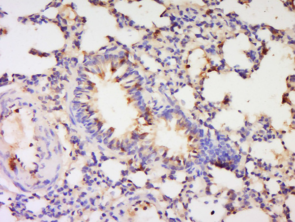
. Visfatin was detected in a paraffin-embedded section of human mammary cancer tissue. Heat mediated antigen retrieval was performed in EDTA buffer (pH 8.0, epitope retrieval solution). The tissue section was blocked with 10% goat serum. The tissue section was then incubated with 1 microg/ml rabbit anti-Visfatin Antibody (PB10009) overnight at 4°C. Biotinylated goat anti-rabbit IgG was used as secondary antibody and incubated for 30 minutes at 37°C. The tissue section was developed using Strepavidin-Biotin-Complex (SABC) (Catalog # SA1022) with DAB as the chromogen.")
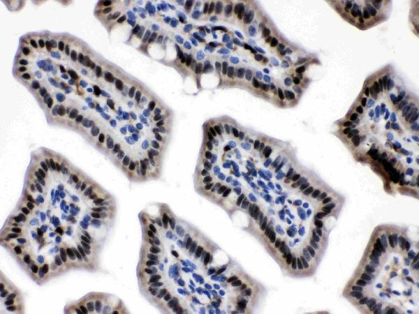
. Visfatin was detected in a paraffin-embedded section of rat intestine tissue. Heat mediated antigen retrieval was performed in EDTA buffer (pH 8.0, epitope retrieval solution). The tissue section was blocked with 10% goat serum. The tissue section was then incubated with 1 microg/ml rabbit anti-Visfatin Antibody (PB10009) overnight at 4°C. Biotinylated goat anti-rabbit IgG was used as secondary antibody and incubated for 30 minutes at 37°C. The tissue section was developed using Strepavidin-Biotin-Complex (SABC) (Catalog # SA1022) with DAB as the chromogen.")
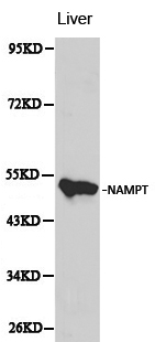
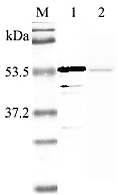
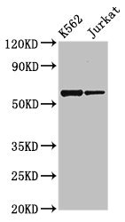
. Electrophoresis was performed on a 5-20% SDS-PAGE gel at 70V (Stacking gel) / 90V (Resolving gel) for 2-3 hours. The sample well of each lane was loaded with 30 ug of sample under reducing conditions. Lane 1: human U87 whole cell lysates, Lane 2: human A431 whole cell lysates, Lane 3: human Daudi whole cell lysates, Lane 4: human U20S whole cell lysates, Lane 5: human K562 whole cell lysates, Lane 6: human A549 whole cell lysates, Lane 7: human HL-60 whole cell lysates, Lane 8: mouse testis tissue lysates, Lane 9: mouse RAW264.7 whole cell lysates, Lane 10: rat brain tissue lysates. After electrophoresis, proteins were transferred to a nitrocellulose membrane at 150 mA for 50-90 minutes. Blocked the membrane with 5% non-fat milk/TBS for 1.5 hour at RT. The membrane was incubated with rabbit anti-Visfatin antigen affinity purified polyclonal antibody (Catalog # PB10009) at 0.5 microg/mL overnight at 4°C, then washed with TBS-0.1%Tween 3 times with 5 minutes each and probed with a goat anti-rabbit IgG-HRP secondary antibody at a dilution of 1:5000 for 1.5 hour at RT. The signal is developed using an Enhanced Chemiluminescent detection (ECL) kit (Catalog # EK1002) with Tanon 5200 system. A specific band was detected for Visfatin at approximately 54 kDa. The expected band size for Visfatin is at 56 kDa.")
. Overlay histogram showing U20S cells stained with PB10009 (Blue line). To facilitate intracellular staining, cells were fixed with 4% paraformaldehyde and permeabilized with permeabilization buffer. The cells were blocked with 10% normal goat serum. And then incubated with rabbit anti-Visfatin Antibody (PB10009, 1 microg/1x106 cells) for 30 min at 20°C. DyLight®488 conjugated goat anti-rabbit IgG (BA1127, 5-10 microg/1x106 cells) was used as secondary antibody for 30 minutes at 20°C. Isotype control antibody (Green line) was rabbit IgG (1 microg/1x106) used under the same conditions. Unlabelled sample without incubation with primary antibody and secondary antibody (Red line) was used as a blank control.")
. Visfatin/NAMPT was detected in an immunocytochemical section of A549 cells. Enzyme antigen retrieval was performed using IHC enzyme antigen retrieval reagent (AR0022) for 15 mins. The cells were blocked with 10% goat serum. And then incubated with 5 microg/mL rabbit anti-Visfatin/NAMPT Antibody (PB10009) overnight at 4°C. DyLight®488 Conjugated Goat Anti-Rabbit IgG (BA1127) was used as secondary antibody at 1:500 dilution and incubated for 30 minutes at 37°C. The section was counterstained with DAPI. Visualize using a fluorescence microscope and filter sets appropriate for the label used.")
Figure 2. IHC analysis of Visfatin using anti-Visfatin antibody (PB10009). Visfatin was detected in a paraffin-embedded section of mouse intestine tissue. Heat mediated antigen retrieval was performed in EDTA buffer (pH 8.0, epitope retrieval solution). The tissue section was blocked with 10% goat serum. The tissue section was then incubated with 1 microg/ml rabbit anti-Visfatin Antibody (PB10009) overnight at 4°C. Biotinylated goat anti-rabbit IgG was used as secondary antibody and incubated for 30 minutes at 37°C. The tissue section was developed using Strepavidin-Biotin-Complex (SABC) (Catalog # SA1022) with DAB as the chromogen.
Anti-Visfatin/NAMPT Antibody Picoband(r)
PB10009-CARRIER-FREE
ApplicationsFlow Cytometry, ImmunoFluorescence, Western Blot, ELISA, ImmunoCytoChemistry, ImmunoHistoChemistry, ImmunoHistoChemistry Frozen
Product group Antibodies
ReactivityHuman, Mouse, Rat
TargetNAMPT
Overview
- SupplierBoster Bio
- Product NameAnti-Visfatin/NAMPT Antibody Picoband(r)
- Delivery Days Customer9
- Application Supplier NoteTested Species: In-house tested species with positive results. By Heat: Boiling the paraffin sections in 10mM citrate buffer, pH6.0, for 20mins is required for the staining of formalin/paraffin sections. Other applications have not been tested. Optimal dilutions should be determined by end users.
- ApplicationsFlow Cytometry, ImmunoFluorescence, Western Blot, ELISA, ImmunoCytoChemistry, ImmunoHistoChemistry, ImmunoHistoChemistry Frozen
- CertificationResearch Use Only
- ClonalityPolyclonal
- Concentration500 ug/ml
- Gene ID10135
- Target nameNAMPT
- Target descriptionnicotinamide phosphoribosyltransferase
- Target synonyms1110035O14Rik, PBEF, PBEF1, VF, VISFATIN, nicotinamide phosphoribosyltransferase, NAmPRTase, pre-B cell-enhancing factor, pre-B-cell colony-enhancing factor 1
- HostRabbit
- IsotypeIgG
- Protein IDP43490
- Protein NameNicotinamide phosphoribosyltransferase
- Scientific DescriptionBoster Bio Anti-Visfatin/NAMPT Antibody Picoband® catalog # PB10009. Tested in ELISA, Flow Cytometry, IF, IHC, IHC-F, ICC, WB applications. This antibody reacts with Human, Mouse, Rat. The brand Picoband indicates this is a premium antibody that guarantees superior quality, high affinity, and strong signals with minimal background in Western blot applications. Only our best-performing antibodies are designated as Picoband, ensuring unmatched performance.
- ReactivityHuman, Mouse, Rat
- Storage Instruction-20°C,2°C to 8°C
- UNSPSC12352203
Related products
Product group Antibodies
Anti-NAMPT AntibodyA28625
ApplicationsImmunoFluorescence, Western Blot, ImmunoHistoChemistry
ReactivityHuman, Mouse, Rat
- SizePrice
Product group Antibodies
anti-Nampt (Visfatin/PBEF) (human), pAbAG-25A-0025
ApplicationsWestern Blot, ELISA, ImmunoHistoChemistry
ReactivityHuman
TargetNAMPT
- SizePrice
Product group Antibodies
Visfatin Polyclonal AntibodyBS-0272R
ApplicationsImmunoFluorescence, Western Blot, ImmunoCytoChemistry, ImmunoHistoChemistry, ImmunoHistoChemistry Frozen, ImmunoHistoChemistry Paraffin
ReactivityBovine, Chicken, Equine, Human, Monkey, Mouse, Porcine, Rat
TargetNAMPT
- SizePrice
Product group Antibodies
NAMPT Polyclonal AntibodyCAC14857
ApplicationsImmunoFluorescence, Western Blot, ELISA, ImmunoHistoChemistry
TargetNAMPT
- SizePrice
Product group Antibodies
NAMPT AntibodyCSB-PA015422EA01HU
ApplicationsImmunoFluorescence, Western Blot, ELISA, ImmunoHistoChemistry
ReactivityHuman
TargetNAMPT
- SizePrice
Product group Antibodies

NAMPT / Visfatin AntibodyLS-C149234
ApplicationsWestern Blot, ELISA
ReactivityHuman, Mouse, Rat
TargetNAMPT
- SizePrice
Product group Antibodies
Visfatin antibody [N1N3]GTX117444
ApplicationsWestern Blot, ImmunoHistoChemistry, ImmunoHistoChemistry Paraffin
ReactivityHuman, Rat
TargetNAMPT
- SizePrice
Product group Antibodies
Anti-Visfatin AntibodyRB-08-0003
ApplicationsWestern Blot, ELISA
ReactivityHuman
- SizePrice