


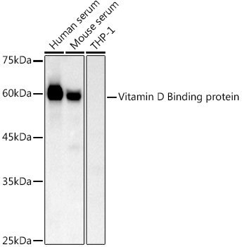
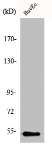
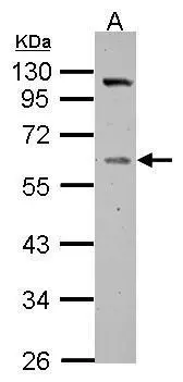
Figure 1. Western blot analysis of Vitamin D Binding protein using anti-Vitamin D Binding protein antibody (A03364-1). Electrophoresis was performed on a 5-20% SDS-PAGE gel at 70V (Stacking gel) / 90V (Resolving gel) for 2-3 hours. The sample well of each lane was loaded with 50ug of sample under reducing conditions. Lane 1: human placenta tissue lysates, Lane 2: human A431 whole cell lysates. After Electrophoresis, proteins were transferred to a Nitrocellulose membrane at 150mA for 50-90 minutes. Blocked the membrane with 5% Non-fat Milk/ TBS for 1.5 hour at RT. The membrane was incubated with rabbit anti-Vitamin D Binding protein antigen affinity purified polyclonal antibody (Catalog # A03364-1) at 0.5 ug/mL overnight at 4 then washed with TBS-0.1%Tween 3 times with 5 minutes each and probed with a goat anti-rabbit IgG-HRP secondary antibody at a dilution of 1:10000 for 1.5 hour at RT. The signal is developed using an Enhanced Chemiluminescent detection (ECL) kit (Catalog # EK1002) with Tanon 5200 system. A specific band was detected for Vitamin D Binding protein at approximately 53KD. The expected band size for Vitamin D Binding protein is at 53KD.
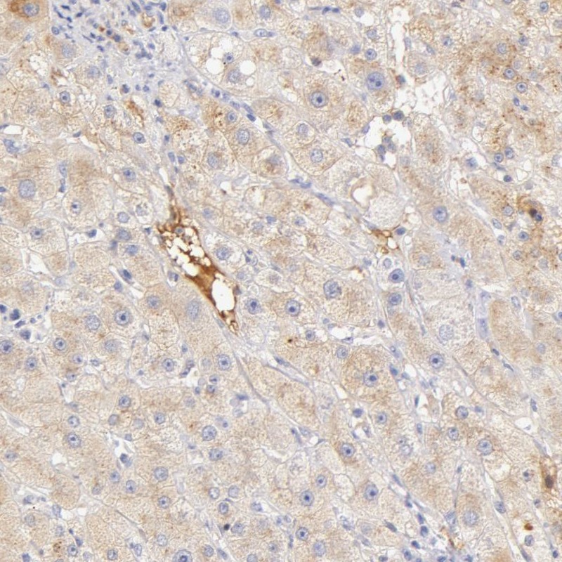
. Vitamin D Binding protein was detected in paraffin-embedded section of human liver cancer tissue. Heat mediated antigen retrieval was performed in citrate buffer (pH6, epitope retrieval solution) for 20 mins. The tissue section was blocked with 10% goat serum. The tissue section was then incubated with 1ug/ml rabbit anti-Vitamin D Binding protein Antibody (A03364-1) overnight at 4 Biotinylated goat anti-rabbit IgG was used as secondary antibody and incubated for 30 minutes at 37 The tissue section was developed using Strepavidin-Biotin-Complex (SABC)(Catalog # SA1022) with DAB as the chromogen.")
. Vitamin D Binding protein was detected in paraffin-embedded section of human lung cancer tissue. Heat mediated antigen retrieval was performed in citrate buffer (pH6, epitope retrieval solution) for 20 mins. The tissue section was blocked with 10% goat serum. The tissue section was then incubated with 1ug/ml rabbit anti-Vitamin D Binding protein Antibody (A03364-1) overnight at 4 Biotinylated goat anti-rabbit IgG was used as secondary antibody and incubated for 30 minutes at 37 The tissue section was developed using Strepavidin-Biotin-Complex (SABC)(Catalog # SA1022) with DAB as the chromogen.")
. Vitamin D Binding protein was detected in paraffin-embedded section of mouse liver tissue. Heat mediated antigen retrieval was performed in citrate buffer (pH6, epitope retrieval solution) for 20 mins. The tissue section was blocked with 10% goat serum. The tissue section was then incubated with 1ug/ml rabbit anti-Vitamin D Binding protein Antibody (A03364-1) overnight at 4 Biotinylated goat anti-rabbit IgG was used as secondary antibody and incubated for 30 minutes at 37 The tissue section was developed using Strepavidin-Biotin-Complex (SABC)(Catalog # SA1022) with DAB as the chromogen.")
. Vitamin D Binding protein was detected in paraffin-embedded section of human rectal cancer tissue. Heat mediated antigen retrieval was performed in citrate buffer (pH6, epitope retrieval solution) for 20 mins. The tissue section was blocked with 10% goat serum. The tissue section was then incubated with 1ug/ml rabbit anti-Vitamin D Binding protein Antibody (A03364-1) overnight at 4 Biotinylated goat anti-rabbit IgG was used as secondary antibody and incubated for 30 minutes at 37 The tissue section was developed using Strepavidin-Biotin-Complex (SABC)(Catalog # SA1022) with DAB as the chromogen.")
Figure 1. Western blot analysis of Vitamin D Binding protein using anti-Vitamin D Binding protein antibody (A03364-1). Electrophoresis was performed on a 5-20% SDS-PAGE gel at 70V (Stacking gel) / 90V (Resolving gel) for 2-3 hours. The sample well of each lane was loaded with 50ug of sample under reducing conditions. Lane 1: human placenta tissue lysates, Lane 2: human A431 whole cell lysates. After Electrophoresis, proteins were transferred to a Nitrocellulose membrane at 150mA for 50-90 minutes. Blocked the membrane with 5% Non-fat Milk/ TBS for 1.5 hour at RT. The membrane was incubated with rabbit anti-Vitamin D Binding protein antigen affinity purified polyclonal antibody (Catalog # A03364-1) at 0.5 ug/mL overnight at 4 then washed with TBS-0.1%Tween 3 times with 5 minutes each and probed with a goat anti-rabbit IgG-HRP secondary antibody at a dilution of 1:10000 for 1.5 hour at RT. The signal is developed using an Enhanced Chemiluminescent detection (ECL) kit (Catalog # EK1002) with Tanon 5200 system. A specific band was detected for Vitamin D Binding protein at approximately 53KD. The expected band size for Vitamin D Binding protein is at 53KD.
Anti-Vitamin D Binding protein/GC Antibody Picoband(r)
A03364-1-CARRIER-FREE
ApplicationsWestern Blot, ELISA, ImmunoHistoChemistry
Product group Antibodies
ReactivityHuman, Mouse, Rat
TargetGC
Overview
- SupplierBoster Bio
- Product NameAnti-Vitamin D Binding protein/GC Antibody Picoband(r)
- Delivery Days Customer9
- ApplicationsWestern Blot, ELISA, ImmunoHistoChemistry
- CertificationResearch Use Only
- ClonalityPolyclonal
- Concentration500 ug/ml
- Gene ID2638
- Target nameGC
- Target descriptionGC vitamin D binding protein
- Target synonymsDBP, DBP-maf, DBP/GC, GRD3, Gc-MAF, GcMAF, HEL-S-51, VDB, VDBG, VDBP, vitamin D-binding protein, epididymis secretory protein Li 51, gc protein-derived macrophage activating factor, gc-globulin, group-specific component (vitamin D binding protein), vitamin D-binding alpha-globulin, vitamin D-binding protein-macrophage activating factor
- HostRabbit
- IsotypeIgG
- Protein IDP02774
- Protein NameVitamin D-binding protein
- Scientific DescriptionBoster Bio Anti-Vitamin D Binding protein/GC Antibody Picoband® catalog # A03364-1. Tested in ELISA, IHC, WB applications. This antibody reacts with Human, Mouse, Rat. The brand Picoband indicates this is a premium antibody that guarantees superior quality, high affinity, and strong signals with minimal background in Western blot applications. Only our best-performing antibodies are designated as Picoband, ensuring unmatched performance.
- ReactivityHuman, Mouse, Rat
- Storage Instruction-20°C,2°C to 8°C
- UNSPSC12352203
Related products
Product group Antibodies
Anti-GC Antibody144-05709
ApplicationsImmunoFluorescence, Western Blot
ReactivityHuman, Mouse, Rat
TargetGC
- SizePrice
Product group Antibodies
ApplicationsWestern Blot
ReactivityHuman, Mouse
- SizePrice
Product group Antibodies
ApplicationsFlow Cytometry, Western Blot
ReactivityHuman
TargetGC
- SizePrice
Product group Antibodies
GC AntibodyCSB-PA002082
ApplicationsWestern Blot, ELISA
ReactivityHuman, Monkey
TargetGC
- SizePrice
Product group Antibodies

ApplicationsWestern Blot, ELISA
ReactivityHuman
TargetGC
- SizePrice
Product group Antibodies
Gc Polyclonal AntibodyCAC07536
ApplicationsWestern Blot, ELISA, ImmunoHistoChemistry
TargetGC
- SizePrice
Product group Antibodies

GC / Vitamin D-Binding Protein AntibodyLS-C401124
ApplicationsWestern Blot, ELISA, ImmunoHistoChemistry
ReactivityHuman
TargetGC
- SizePrice
Product group Antibodies
Anti-GC AntibodyHPA001526
ApplicationsWestern Blot, ImmunoHistoChemistry
ReactivityHuman
TargetGC
- SizePrice
Product group Antibodies
Vitamin D Binding Protein antibodyGTX109955
ApplicationsImmunoFluorescence, Western Blot, ImmunoCytoChemistry, ImmunoHistoChemistry, ImmunoHistoChemistry Paraffin
ReactivityHuman, Mouse
TargetGC
- SizePrice
Product group Antibodies
TargetGC
- SizePrice