


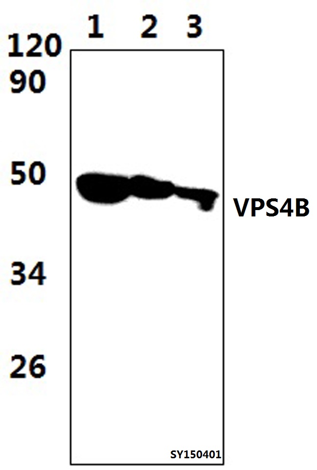
Figure 1. Western blot analysis of VPS4B/MIG1 using anti-VPS4B/MIG1 antibody (A03403-1). Electrophoresis was performed on a 5-20% SDS-PAGE gel at 70V (Stacking gel) / 90V (Resolving gel) for 2-3 hours. The sample well of each lane was loaded with 50ug of sample under reducing conditions. Lane 1: human Hela whole cell lysates, Lane 2: human HEK293 whole cell lysates, Lane 3: human Jurkat whole cell lysates, Lane 4: human SKOV-3 whole cell lysates, Lane 5: rat kidney tissue lysates, Lane 6: rat PC-12 whole cell lysates, Lane 7: mouse brain tissue lysates, Lane 8: mouse NIH/3T3 whole cell lysates, After Electrophoresis, proteins were transferred to a Nitrocellulose membrane at 150mA for 50-90 minutes. Blocked the membrane with 5% Non-fat Milk/ TBS for 1.5 hour at RT. The membrane was incubated with rabbit anti-VPS4B/MIG1 antigen affinity purified polyclonal antibody (Catalog # A03403-1) at 0.5 microg/mL overnight at 4°C, then washed with TBS-0.1%Tween 3 times with 5 minutes each and probed with a goat anti-rabbit IgG-HRP secondary antibody at a dilution of 1:10000 for 1.5 hour at RT. The signal is developed using an Enhanced Chemiluminescent detection (ECL) kit (Catalog # EK1002) with Tanon 5200 system. A specific band was detected for VPS4B/MIG1 at approximately 50KD. The expected band size for VPS4B/MIG1 is at 50KD.
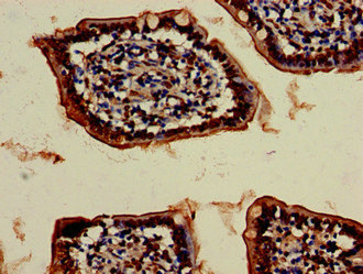
. VPS4B/MIG1 was detected in paraffin-embedded section of human lung cancer tissue. Heat mediated antigen retrieval was performed in EDTA buffer (pH8.0, epitope retrieval solution). The tissue section was blocked with 10% goat serum. The tissue section was then incubated with 1microg/ml rabbit anti-VPS4B/MIG1 Antibody (A03403-1) overnight at 4°C. Biotinylated goat anti-rabbit IgG was used as secondary antibody and incubated for 30 minutes at 37°C. The tissue section was developed using Strepavidin-Biotin-Complex (SABC) (Catalog # SA1022) with DAB as the chromogen.")
. VPS4B/MIG1 was detected in paraffin-embedded section of human lung cancer tissue. Heat mediated antigen retrieval was performed in EDTA buffer (pH8.0, epitope retrieval solution). The tissue section was blocked with 10% goat serum. The tissue section was then incubated with 1microg/ml rabbit anti-VPS4B/MIG1 Antibody (A03403-1) overnight at 4°C. Biotinylated goat anti-rabbit IgG was used as secondary antibody and incubated for 30 minutes at 37°C. The tissue section was developed using Strepavidin-Biotin-Complex (SABC) (Catalog # SA1022) with DAB as the chromogen.")
. Overlay histogram showing A431 cells stained with A03403-1 (Blue line). To facilitate intracellular staining, cells were fixed with 4% paraformaldehyde and permeabilized with permeabilization buffer. The cells were blocked with 10% normal goat serum. And then incubated with rabbit anti-VPS4B/MIG1 Antibody (A03403-1,1microg/1x106 cells) for 30 min at 20°C. DyLight®488 conjugated goat anti-rabbit IgG (BA1127, 5-10microg/1x106 cells) was used as secondary antibody for 30 minutes at 20°C. Isotype control antibody (Green line) was rabbit IgG (1microg/1x106) used under the same conditions. Unlabelled sample without incubation with primary antibody and secondary antibody (Red line) was used as a blank control.")
Figure 1. Western blot analysis of VPS4B/MIG1 using anti-VPS4B/MIG1 antibody (A03403-1). Electrophoresis was performed on a 5-20% SDS-PAGE gel at 70V (Stacking gel) / 90V (Resolving gel) for 2-3 hours. The sample well of each lane was loaded with 50ug of sample under reducing conditions. Lane 1: human Hela whole cell lysates, Lane 2: human HEK293 whole cell lysates, Lane 3: human Jurkat whole cell lysates, Lane 4: human SKOV-3 whole cell lysates, Lane 5: rat kidney tissue lysates, Lane 6: rat PC-12 whole cell lysates, Lane 7: mouse brain tissue lysates, Lane 8: mouse NIH/3T3 whole cell lysates, After Electrophoresis, proteins were transferred to a Nitrocellulose membrane at 150mA for 50-90 minutes. Blocked the membrane with 5% Non-fat Milk/ TBS for 1.5 hour at RT. The membrane was incubated with rabbit anti-VPS4B/MIG1 antigen affinity purified polyclonal antibody (Catalog # A03403-1) at 0.5 microg/mL overnight at 4°C, then washed with TBS-0.1%Tween 3 times with 5 minutes each and probed with a goat anti-rabbit IgG-HRP secondary antibody at a dilution of 1:10000 for 1.5 hour at RT. The signal is developed using an Enhanced Chemiluminescent detection (ECL) kit (Catalog # EK1002) with Tanon 5200 system. A specific band was detected for VPS4B/MIG1 at approximately 50KD. The expected band size for VPS4B/MIG1 is at 50KD.
Anti-VPS4B/MIG1 Antibody Picoband(r)
A03403-1-CARRIER-FREE
ApplicationsFlow Cytometry, Western Blot, ELISA, ImmunoHistoChemistry
Product group Antibodies
ReactivityHuman, Mouse, Rat
TargetVPS4B
Overview
- SupplierBoster Bio
- Product NameAnti-VPS4B/MIG1 Antibody Picoband(r)
- Delivery Days Customer9
- ApplicationsFlow Cytometry, Western Blot, ELISA, ImmunoHistoChemistry
- CertificationResearch Use Only
- ClonalityPolyclonal
- Concentration500 ug/ml
- Gene ID9525
- Target nameVPS4B
- Target descriptionvacuolar protein sorting 4 homolog B
- Target synonymsMIG1, SKD1, SKD1B, VPS4-2, vacuolar protein sorting-associated protein 4B, cell migration-inducing 1, cell migration-inducing gene 1 protein, suppressor of K(+) transport growth defect 1, suppressor of K+ transport defect 1, vacuolar protein sorting 4B
- HostRabbit
- IsotypeIgG
- Protein IDO75351
- Protein NameVacuolar protein sorting-associated protein 4B
- Scientific DescriptionBoster Bio Anti-VPS4B/MIG1 Antibody Picoband® catalog # A03403-1. Tested in ELISA, Flow Cytometry, IHC, WB applications. This antibody reacts with Human, Mouse, Rat. The brand Picoband indicates this is a premium antibody that guarantees superior quality, high affinity, and strong signals with minimal background in Western blot applications. Only our best-performing antibodies are designated as Picoband, ensuring unmatched performance.
- ReactivityHuman, Mouse, Rat
- Storage Instruction-20°C,2°C to 8°C
- UNSPSC12352203
Related products
Product group Antibodies
Anti-VPS4B AntibodyA29314
ApplicationsWestern Blot
ReactivityHuman, Mouse, Rat
- SizePrice
Product group Antibodies

SKD1 / VPS4B Antibody (Biotin)LS-C680003
ApplicationsELISA
ReactivityHuman
TargetVPS4B
- SizePrice
Product group Antibodies
References
VPS4B Polyclonal AntibodyBS-12778R
ApplicationsImmunoFluorescence, Western Blot, ELISA, ImmunoCytoChemistry, ImmunoHistoChemistry, ImmunoHistoChemistry Frozen, ImmunoHistoChemistry Paraffin
ReactivityBovine, Fish, Human, Mouse, Porcine, Rat
TargetVPS4B
- SizePrice
Product group Antibodies
Vps4B Polyclonal AntibodyCAC08972
ApplicationsImmunoFluorescence, Western Blot, ELISA, ImmunoHistoChemistry
ReactivityMouse
TargetVPS4B
- SizePrice
Product group Antibodies
VPS4B AntibodyCSB-PA025915LA01HU
ApplicationsImmunoFluorescence, Western Blot, ELISA, ImmunoHistoChemistry
ReactivityHuman, Mouse
TargetVPS4B
- SizePrice

Product group Antibodies
Anti-VPS4B AntibodyHPA057649
ApplicationsImmunoHistoChemistry
ReactivityHuman
TargetVPS4B
- SizePrice
![VPS4B antibody [N3C3] detects VPS4B protein at nucleus and endosome by immunofluorescent analysis. Sample: A375 cells were fixed in 4% paraformaldehyde at RT for 15 min. Green: VPS4B protein stained by VPS4B antibody [N3C3] (GTX115249) diluted at 1:500. Blue: Hoechst 33342 staining. Scale bar = 10 μm.](https://www.genetex.com/upload/website/prouct_img/normal/GTX115249/GTX115249_40254_IFA_2_w_23060519_512.webp)
Product group Antibodies
VPS4B antibody [N3C3]GTX115249
ApplicationsImmunoFluorescence, Western Blot, ImmunoCytoChemistry, ImmunoHistoChemistry, ImmunoHistoChemistry Paraffin
ReactivityHuman
TargetVPS4B
- SizePrice
Product group Antibodies
VPS4B AntibodyPACO20863
ApplicationsELISA, ImmunoHistoChemistry
ReactivityHuman
TargetVPS4B
- SizePrice