

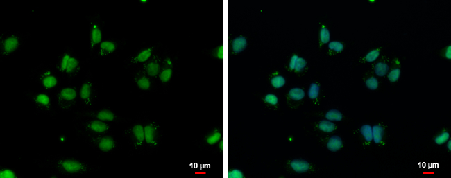
VPS4B antibody [N3C3] detects VPS4B protein at nucleus and endosome by immunofluorescent analysis. Sample: A375 cells were fixed in 4% paraformaldehyde at RT for 15 min. Green: VPS4B protein stained by VPS4B antibody [N3C3] (GTX115249) diluted at 1:500. Blue: Hoechst 33342 staining. Scale bar = 10 μm.
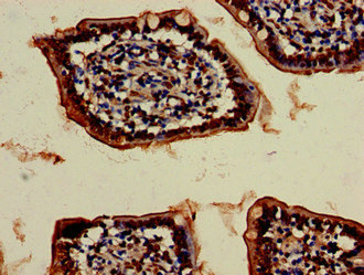
![VPS4B antibody [N3C3] detects VPS4B protein at nucleus on human colon carcinoma by immunohistochemical analysis. Sample: Paraffin-embedded colon carcinoma. VPS4B antibody [N3C3] (GTX115249) dilution: 1:500.
Antigen Retrieval: Trilogy? (EDTA based, pH 8.0) buffer, 15min](https://www.genetex.com/upload/website/prouct_img/normal/GTX115249/GTX115249_40254_IHC_w_23060519_213.webp "VPS4B antibody [N3C3] detects VPS4B protein at nucleus on human colon carcinoma by immunohistochemical analysis. Sample: Paraffin-embedded colon carcinoma. VPS4B antibody [N3C3] (GTX115249) dilution: 1:500.
Antigen Retrieval: Trilogy? (EDTA based, pH 8.0) buffer, 15min")
![VPS4B antibody [N3C3] detects VPS4B protein at nucleus by immunofluorescent analysis. Sample: HeLa cells were fixed in 4% paraformaldehyde at RT for 15 min. Green: VPS4B protein stained by VPS4B antibody [N3C3] (GTX115249) diluted at 1:500. Red: alpha Tubulin, a cytoskeleton marker, stained by alpha Tubulin antibody [B-5-1-2] (GTX11304) diluted at 1:10000. Blue: Hoechst 33342 staining.](https://www.genetex.com/upload/website/prouct_img/normal/GTX115249/GTX115249_40254_20150410_IFA_w_23060519_108.webp "VPS4B antibody [N3C3] detects VPS4B protein at nucleus by immunofluorescent analysis. Sample: HeLa cells were fixed in 4% paraformaldehyde at RT for 15 min. Green: VPS4B protein stained by VPS4B antibody [N3C3] (GTX115249) diluted at 1:500. Red: alpha Tubulin, a cytoskeleton marker, stained by alpha Tubulin antibody [B-5-1-2] (GTX11304) diluted at 1:10000. Blue: Hoechst 33342 staining.")
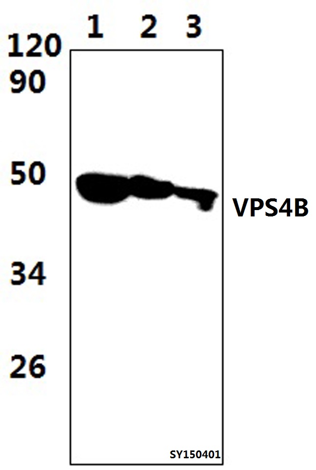
![Various whole cell extracts (30 μg) were separated by 10% SDS-PAGE, and the membrane was blotted with VPS4B antibody [N3C3] (GTX115249) diluted at 1:3000. The HRP-conjugated anti-rabbit IgG antibody (GTX213110-01) was used to detect the primary antibody. Corresponding RNA expression data are based on Human Protein Atlas program.](https://www.genetex.com/upload/website/prouct_img/normal/GTX115249/GTX115249_45708_20250314_WB_TPM_watermark_25032719_703.webp "Various whole cell extracts (30 μg) were separated by 10% SDS-PAGE, and the membrane was blotted with VPS4B antibody [N3C3] (GTX115249) diluted at 1:3000. The HRP-conjugated anti-rabbit IgG antibody (GTX213110-01) was used to detect the primary antibody. Corresponding RNA expression data are based on Human Protein Atlas program.")
VPS4B antibody [N3C3] detects VPS4B protein at nucleus and endosome by immunofluorescent analysis. Sample: A375 cells were fixed in 4% paraformaldehyde at RT for 15 min. Green: VPS4B protein stained by VPS4B antibody [N3C3] (GTX115249) diluted at 1:500. Blue: Hoechst 33342 staining. Scale bar = 10 μm.
VPS4B antibody [N3C3]
GTX115249
ApplicationsImmunoFluorescence, Western Blot, ImmunoCytoChemistry, ImmunoHistoChemistry, ImmunoHistoChemistry Paraffin
Product group Antibodies
ReactivityHuman
TargetVPS4B
Overview
- SupplierGeneTex
- Product NameVPS4B antibody [N3C3]
- Delivery Days Customer9
- Application Supplier NoteWB: 1:500-1:3000. ICC/IF: 1:100-1:1000. IHC-P: 1:100-1:1000. *Optimal dilutions/concentrations should be determined by the researcher.Not tested in other applications.
- ApplicationsImmunoFluorescence, Western Blot, ImmunoCytoChemistry, ImmunoHistoChemistry, ImmunoHistoChemistry Paraffin
- CertificationResearch Use Only
- ClonalityPolyclonal
- Concentration0.32 mg/ml
- ConjugateUnconjugated
- Gene ID9525
- Target nameVPS4B
- Target descriptionvacuolar protein sorting 4 homolog B
- Target synonymsMIG1, SKD1, SKD1B, VPS4-2, vacuolar protein sorting-associated protein 4B, cell migration-inducing 1, cell migration-inducing gene 1 protein, suppressor of K(+) transport growth defect 1, suppressor of K+ transport defect 1, vacuolar protein sorting 4B
- HostRabbit
- IsotypeIgG
- Protein IDO75351
- Protein NameVacuolar protein sorting-associated protein 4B
- Scientific DescriptionThe protein encoded by this gene is a member of the AAA protein family (ATPases associated with diverse cellular activities), and is the homolog of the yeast Vps4 protein. In humans, two paralogs of the yeast protein have been identified. The former share a high degree of aa sequence similarity with each other, and also with yeast Vps4 and mouse Skd1 proteins. Mouse Skd1 (suppressor of K+ transport defect 1) has been shown to be a yeast Vps4 ortholog. Functional studies indicate that both human paralogs associate with the endosomal compartments, and are involved in intracellular protein trafficking, similar to Vps4 protein in yeast. The gene encoding this paralog has been mapped to chromosome 18; the gene for the other resides on chromosome 16. [provided by RefSeq]
- ReactivityHuman
- Storage Instruction-20°C or -80°C,2°C to 8°C
- UNSPSC41116161
Datasheet
Related products
Product group Antibodies
Anti-VPS4B AntibodyA29314
ApplicationsWestern Blot
ReactivityHuman, Mouse, Rat
- SizePrice

Product group Antibodies
Anti-VPS4B/MIG1 Antibody Picoband(r)A03403-1-CARRIER-FREE
ApplicationsFlow Cytometry, Western Blot, ELISA, ImmunoHistoChemistry
ReactivityHuman, Mouse, Rat
TargetVPS4B
- SizePrice
Product group Antibodies

SKD1 / VPS4B Antibody (Biotin)LS-C680003
ApplicationsELISA
ReactivityHuman
TargetVPS4B
- SizePrice
Product group Antibodies
References
VPS4B Polyclonal AntibodyBS-12778R
ApplicationsImmunoFluorescence, Western Blot, ELISA, ImmunoCytoChemistry, ImmunoHistoChemistry, ImmunoHistoChemistry Frozen, ImmunoHistoChemistry Paraffin
ReactivityBovine, Fish, Human, Mouse, Porcine, Rat
TargetVPS4B
- SizePrice
Product group Antibodies
Vps4B Polyclonal AntibodyCAC08972
ApplicationsImmunoFluorescence, Western Blot, ELISA, ImmunoHistoChemistry
ReactivityMouse
TargetVPS4B
- SizePrice
Product group Antibodies
VPS4B AntibodyCSB-PA025915LA01HU
ApplicationsImmunoFluorescence, Western Blot, ELISA, ImmunoHistoChemistry
ReactivityHuman, Mouse
TargetVPS4B
- SizePrice

Product group Antibodies
Anti-VPS4B AntibodyHPA057649
ApplicationsImmunoHistoChemistry
ReactivityHuman
TargetVPS4B
- SizePrice
Product group Antibodies
VPS4B AntibodyPACO20863
ApplicationsELISA, ImmunoHistoChemistry
ReactivityHuman
TargetVPS4B
- SizePrice