


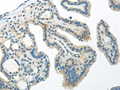
Immunohistochemical staining of human prostate shows strong cytoplasmic positivity in smooth muscle cells.
Immunohistochemical staining of human prostate shows strong cytoplasmic positivity in smooth muscle cells.
Anti-WNT10A Antibody
HPA013898
ApplicationsImmunoHistoChemistry
Product group Antibodies
ReactivityHuman
TargetWNT10A
Overview
- SupplierAtlas Antibodies
- Product NameAnti-WNT10A Antibody
- Delivery Days Customer4
- ApplicationsImmunoHistoChemistry
- CertificationResearch Use Only
- ClonalityPolyclonal
- ConjugateUnconjugated
- Gene ID80326
- Target nameWNT10A
- Target descriptionWnt family member 10A
- Target synonymsECTD16, OODD, SSPS, STHAG4, protein Wnt-10a, wingless-type MMTV integration site family, member 10A
- HostRabbit
- IsotypeIgG
- Protein IDQ9GZT5
- Protein NameProtein Wnt-10a
- Scientific DescriptionRecombinant Protein Epitope Signature Tag (PrEST) antigen sequence
- ReactivityHuman
- Storage Instruction-20°C,2°C to 8°C
- UNSPSC41116161
Datasheet
MSDS
Related products

Product group Antibodies
Anti-Wnt10a Picoband(r) AntibodyA03479-2-CARRIER-FREE
ApplicationsFlow Cytometry, Western Blot, ELISA
ReactivityHuman, Mouse, Rat
TargetWNT10A
- SizePrice
Product group Antibodies
Anti-WNT10A Antibody107-10957
ApplicationsWestern Blot
ReactivityHuman
TargetWNT10A
- SizePrice
Product group Antibodies
Anti-Wnt10a AntibodyA90034
ApplicationsWestern Blot
ReactivityHuman, Mouse, Rat
- SizePrice
Product group Antibodies
WNT10A Polyclonal AntibodyBS-1947R
ApplicationsImmunoFluorescence, ELISA, ImmunoCytoChemistry, ImmunoHistoChemistry, ImmunoHistoChemistry Frozen, ImmunoHistoChemistry Paraffin
ReactivityBovine, Canine, Chicken, Equine, Human, Mouse, Porcine, Rabbit, Rat
TargetWNT10A
- SizePrice
Product group Antibodies
WNT10A AntibodyCSB-PA554204
ApplicationsWestern Blot, ELISA, ImmunoHistoChemistry
ReactivityHuman, Mouse
TargetWNT10A
- SizePrice
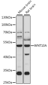
![Whole cell extract (30 μg) was separated by 10% SDS-PAGE, and the membrane was blotted with Wnt10a antibody [N1C1-2] (GTX111191) diluted at 1:1000. The HRP-conjugated anti-rabbit IgG antibody (GTX213110-01) was used to detect the primary antibody, and the signal was developed with Trident ECL plus-Enhanced.](https://www.genetex.com/upload/website/prouct_img/normal/GTX111191/GTX111191_40058_20200529_WB_w_23060500_247.webp)
Product group Antibodies
Wnt10a antibody [N1C1-2]GTX111191
ApplicationsImmunoFluorescence, Western Blot, ImmunoCytoChemistry
ReactivityHuman
TargetWNT10A
- SizePrice
Product group Antibodies

WNT10A AntibodyLS-C749658
ApplicationsWestern Blot
ReactivityHuman
TargetWNT10A
- SizePrice
Product group Antibodies
WNT10A AntibodyPACO20922
ApplicationsELISA, ImmunoHistoChemistry
ReactivityHuman, Mouse
TargetWNT10A
- SizePrice