


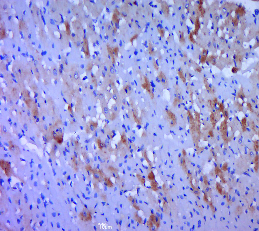
Immunofluorescent staining of human cell line U-2 OS shows localization to vesicles.
Immunofluorescent staining of human cell line U-2 OS shows localization to vesicles.
Anti-WNT2B Antibody
HPA066368
ApplicationsImmunoCytoChemistry
Product group Antibodies
ReactivityHuman
TargetWNT2B
Overview
- SupplierAtlas Antibodies
- Product NameAnti-WNT2B Antibody
- Delivery Days Customer4
- ApplicationsImmunoCytoChemistry
- CertificationResearch Use Only
- ClonalityPolyclonal
- ConjugateUnconjugated
- Gene ID7482
- Target nameWNT2B
- Target descriptionWnt family member 2B
- Target synonymsWNT13, protein Wnt-2b, XWNT2, Xenopus, homolog of, wingless-type MMTV integration site family, member 13, wingless-type MMTV integration site family, member 2B
- HostRabbit
- IsotypeIgG
- Protein IDQ93097
- Protein NameProtein Wnt-2b
- Scientific DescriptionRecombinant Protein Epitope Signature Tag (PrEST) antigen sequence
- ReactivityHuman
- Storage Instruction-20°C,2°C to 8°C
- UNSPSC41116161
Datasheet
MSDS
Related products
Product group Antibodies
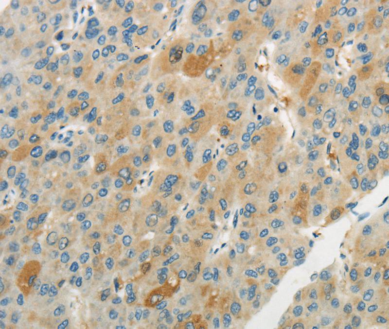
Anti-WNT2B AntibodyA46372
ApplicationsImmunoHistoChemistry
ReactivityHuman
- SizePrice
Product group Antibodies
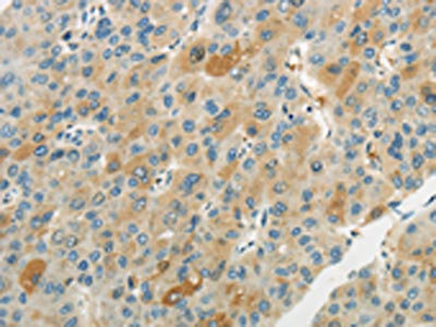
WNT2B AntibodyCSB-PA122866
ApplicationsELISA, ImmunoHistoChemistry
ReactivityHuman, Mouse
TargetWNT2B
- SizePrice

Product group Antibodies
Anti-WNT2B AntibodyHPA047274
ApplicationsImmunoCytoChemistry
ReactivityHuman
TargetWNT2B
- SizePrice
Product group Antibodies
Anti-WNT2B AntibodyHPA047274
ApplicationsImmunoCytoChemistry
ReactivityHuman
TargetWNT2B
- SizePrice

Product group Antibodies
Anti-Wnt2b Antibody Picoband(r)A04879-1-CARRIER-FREE
ApplicationsWestern Blot, ELISA
ReactivityHuman, Mouse, Rat
TargetWNT2B
- SizePrice
Product group Antibodies

WNT2B Antibody (aa169-185)LS-C313116
ApplicationsWestern Blot
ReactivityBovine, Equine, Guinea Pig, Human, Mammals, Monkey, Mouse, Porcine, Rabbit, Rat, Sheep
TargetWNT2B
- SizePrice
Product group Antibodies
ApplicationsImmunoPrecipitation, Western Blot, ImmunoCytoChemistry, ImmunoHistoChemistry
ReactivityMouse, Porcine, Rat
TargetWNT2B
- SizePrice
Product group Antibodies
WNT2B Polyclonal AntibodyBS-1946R
ApplicationsImmunoFluorescence, Western Blot, ELISA, ImmunoCytoChemistry, ImmunoHistoChemistry, ImmunoHistoChemistry Frozen, ImmunoHistoChemistry Paraffin
ReactivityCanine, Chicken, Equine, Human, Mouse, Porcine, Rabbit, Rat
TargetWNT2B
- SizePrice