


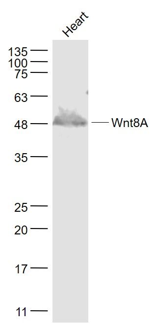
Figure 1. Western blot analysis of Wnt8a using anti-Wnt8a antibody (A08392-2). Electrophoresis was performed on a 5-20% SDS-PAGE gel at 70V (Stacking gel) / 90V (Resolving gel) for 2-3 hours. The sample well of each lane was loaded with 50ug of sample under reducing conditions. Lane 1: human U87 whole cell lysates, Lane 2: human SH-SY5Y whole cell lysates, Lane 3: human SW620 whole cell lysates, Lane 4: human Raji whole cell lysates, Lane 5: rat brain tissue lysates, Lane 6: rat heart tissue lysates, Lane 7: rat C6 whole cell lysates, Lane 8: mouse brain tissue lysates, Lane 9: mouse Neuro-2a whole cell lysates. After Electrophoresis, proteins were transferred to a Nitrocellulose membrane at 150mA for 50-90 minutes. Blocked the membrane with 5% Non-fat Milk/ TBS for 1.5 hour at RT. The membrane was incubated with rabbit anti-Wnt8a antigen affinity purified polyclonal antibody (Catalog # A08392-2) at 0.5 microg/mL overnight at 4°C, then washed with TBS-0.1%Tween 3 times with 5 minutes each and probed with a goat anti-rabbit IgG-HRP secondary antibody at a dilution of 1:5000 for 1.5 hour at RT. The signal is developed using an Enhanced Chemiluminescent detection (ECL) kit (Catalog # EK1002) with Tanon 5200 system. A specific band was detected for Wnt8a at approximately 60KD. The expected band size for Wnt8a is at 60KD.
Figure 1. Western blot analysis of Wnt8a using anti-Wnt8a antibody (A08392-2). Electrophoresis was performed on a 5-20% SDS-PAGE gel at 70V (Stacking gel) / 90V (Resolving gel) for 2-3 hours. The sample well of each lane was loaded with 50ug of sample under reducing conditions. Lane 1: human U87 whole cell lysates, Lane 2: human SH-SY5Y whole cell lysates, Lane 3: human SW620 whole cell lysates, Lane 4: human Raji whole cell lysates, Lane 5: rat brain tissue lysates, Lane 6: rat heart tissue lysates, Lane 7: rat C6 whole cell lysates, Lane 8: mouse brain tissue lysates, Lane 9: mouse Neuro-2a whole cell lysates. After Electrophoresis, proteins were transferred to a Nitrocellulose membrane at 150mA for 50-90 minutes. Blocked the membrane with 5% Non-fat Milk/ TBS for 1.5 hour at RT. The membrane was incubated with rabbit anti-Wnt8a antigen affinity purified polyclonal antibody (Catalog # A08392-2) at 0.5 microg/mL overnight at 4°C, then washed with TBS-0.1%Tween 3 times with 5 minutes each and probed with a goat anti-rabbit IgG-HRP secondary antibody at a dilution of 1:5000 for 1.5 hour at RT. The signal is developed using an Enhanced Chemiluminescent detection (ECL) kit (Catalog # EK1002) with Tanon 5200 system. A specific band was detected for Wnt8a at approximately 60KD. The expected band size for Wnt8a is at 60KD.
Anti-Wnt8a Antibody Picoband(r)
A08392-2-CARRIER-FREE
ApplicationsWestern Blot, ELISA
Product group Antibodies
ReactivityHuman, Mouse, Rat
TargetWNT8A
Overview
- SupplierBoster Bio
- Product NameAnti-Wnt8a Antibody Picoband(r)
- Delivery Days Customer9
- ApplicationsWestern Blot, ELISA
- CertificationResearch Use Only
- ClonalityPolyclonal
- Concentration500 ug/ml
- Gene ID7478
- Target nameWNT8A
- Target descriptionWnt family member 8A
- Target synonymsWNT8D, protein Wnt-8a, WNT8d, protein Wnt-8d, wingless-type MMTV integration site family, member 8A
- HostRabbit
- IsotypeIgG
- Protein IDQ9H1J5
- Protein NameProtein Wnt-8a
- Scientific DescriptionBoster Bio Anti-Wnt8a Antibody Picoband® catalog # A08392-2. Tested in ELISA, WB applications. This antibody reacts with Human, Mouse, Rat. The brand Picoband indicates this is a premium antibody that guarantees superior quality, high affinity, and strong signals with minimal background in Western blot applications. Only our best-performing antibodies are designated as Picoband, ensuring unmatched performance.
- ReactivityHuman, Mouse, Rat
- Storage Instruction-20°C,2°C to 8°C
- UNSPSC12352203
Related products
Product group Antibodies
Anti-WNT8A AntibodyA46479
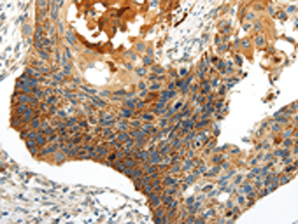
ApplicationsImmunoHistoChemistry
ReactivityHuman
- SizePrice
Product group Antibodies
Wnt8A Polyclonal AntibodyBS-6129R
ApplicationsImmunoFluorescence, Western Blot, ELISA, ImmunoCytoChemistry, ImmunoHistoChemistry, ImmunoHistoChemistry Frozen, ImmunoHistoChemistry Paraffin
ReactivityCanine, Equine, Human, Mouse, Rat, Sheep
TargetWNT8A
- SizePrice
Product group Antibodies
WNT8A AntibodyCSB-PA863951LA01HU
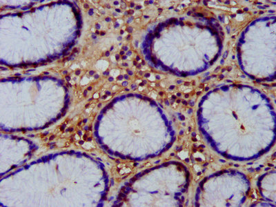
ApplicationsELISA, ImmunoHistoChemistry
ReactivityHuman
TargetWNT8A
- SizePrice
Product group Antibodies

WNT8A Antibody (aa23-358)LS-C371067
ApplicationsELISA
ReactivityXenopus
TargetWNT8A
- SizePrice
Product group Antibodies
Wnt8a antibodyGTX128108
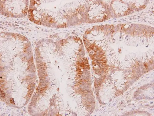
ApplicationsWestern Blot, ImmunoHistoChemistry, ImmunoHistoChemistry Paraffin
ReactivityHuman
TargetWNT8A
- SizePrice
Product group Antibodies
Anti-WNT8A AntibodyHPA038539
ApplicationsWestern Blot, ImmunoHistoChemistry
ReactivityHuman
TargetWNT8A
- SizePrice
Product group Antibodies
WNT8A AntibodyPACO20919
ApplicationsELISA, ImmunoHistoChemistry
ReactivityHuman
TargetWNT8A
- SizePrice