


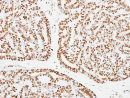
Immunohistochemical staining of human kidney shows moderate to strong nuclear positivity in cells in tubules.
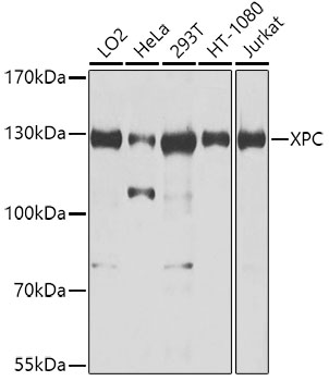
![Lane 1: Marker [kDa] 230, 130, 95, 72, 56, 36, 28, 17, 11. Lane 2: Human cell line RT-4. Lane 3: Human cell line U-251 MG](https://atlasantibodies.s3.amazonaws.com/images/wb/hpa035707-wb-1.jpg "Lane 1: Marker [kDa] 230, 130, 95, 72, 56, 36, 28, 17, 11. Lane 2: Human cell line RT-4. Lane 3: Human cell line U-251 MG")
Immunohistochemical staining of human kidney shows moderate to strong nuclear positivity in cells in tubules.
Anti-XPC Antibody
HPA035707
ApplicationsWestern Blot, ImmunoCytoChemistry, ImmunoHistoChemistry
Product group Antibodies
ReactivityHuman
TargetXPC
Overview
- SupplierAtlas Antibodies
- Product NameAnti-XPC Antibody
- Delivery Days Customer4
- ApplicationsWestern Blot, ImmunoCytoChemistry, ImmunoHistoChemistry
- CertificationResearch Use Only
- ClonalityPolyclonal
- ConjugateUnconjugated
- Gene ID7508
- Target nameXPC
- Target descriptionXPC complex subunit, DNA damage recognition and repair factor
- Target synonymsRAD4, XP3, XPCC, p125, DNA repair protein complementing XP-C cells, mutant xeroderma pigmentosum group C, xeroderma pigmentosum, complementation group C
- HostRabbit
- IsotypeIgG
- Protein IDQ01831
- Protein NameDNA repair protein complementing XP-C cells
- Scientific DescriptionRecombinant Protein Epitope Signature Tag (PrEST) antigen sequence
- ReactivityHuman
- Storage Instruction-20°C,2°C to 8°C
- UNSPSC41116161
Datasheet
MSDS
Related products
Product group Antibodies
Anti-XPC AntibodyA10400
ApplicationsImmunoPrecipitation, Western Blot, ImmunoHistoChemistry
ReactivityHuman
- SizePrice
Product group Antibodies
Anti-XPC Antibody144-08354
ApplicationsImmunoPrecipitation, Western Blot, ImmunoHistoChemistry
ReactivityHuman
TargetXPC
- SizePrice
Product group Antibodies

XPC AntibodyLS-C832647
ApplicationsELISA, ImmunoHistoChemistry
ReactivityHuman
TargetXPC
- SizePrice

Product group Antibodies
Anti-XPC Antibody Picoband(r)A00473-1-CARRIER-FREE
ApplicationsFlow Cytometry, ImmunoFluorescence, Western Blot, ELISA, ImmunoCytoChemistry
ReactivityHuman, Mouse, Rat
TargetXPC
- SizePrice
Product group Antibodies
XPC Polyclonal AntibodyBS-25269R
ApplicationsWestern Blot, ELISA
ReactivityBovine, Equine, Human, Mouse, Porcine, Rat
TargetXPC
- SizePrice
Product group Antibodies
XPC AntibodyCSB-PA026217LA01HU
ApplicationsELISA
ReactivityHuman
TargetXPC
- SizePrice
Product group Antibodies
XPC antibody [C2C3], C-termGTX102840
ApplicationsWestern Blot, ImmunoHistoChemistry, ImmunoHistoChemistry Paraffin
ReactivityHuman
TargetXPC
- SizePrice

Product group Antibodies
Anti-XPC AntibodyHPA069673
ApplicationsImmunoCytoChemistry
ReactivityHuman
TargetXPC
- SizePrice