


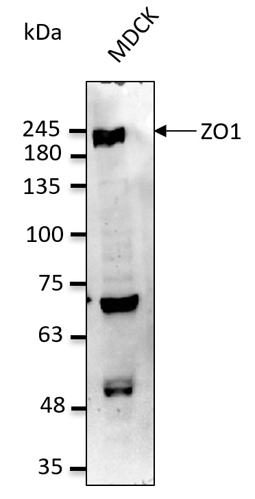
Figure 1. Western blot analysis of TJP1 using anti-TJP1 antibody (PB9234). Electrophoresis was performed on a 5-20% SDS-PAGE gel at 70V (Stacking gel) / 90V (Resolving gel) for 2-3 hours. The sample well of each lane was loaded with 30 ug of sample under reducing conditions. Lane 1: human PC-3 whole cell lysates, Lane 2: human CACO-2 whole cell lysates, Lane 3: human COLO320 whole cell lysates, Lane 4: rat testis tissue lysates, Lane 5: mouse testis tissue lysates. After electrophoresis, proteins were transferred to a nitrocellulose membrane at 150 mA for 50-90 minutes. Blocked the membrane with 5% non-fat milk/TBS for 1.5 hour at RT. The membrane was incubated with rabbit anti-TJP1 antigen affinity purified polyclonal antibody (Catalog # PB9234) at 0.5 microg/mL overnight at 4°C, then washed with TBS-0.1%Tween 3 times with 5 minutes each and probed with a goat anti-rabbit IgG-HRP secondary antibody at a dilution of 1:5000 for 1.5 hour at RT. The signal is developed using an Enhanced Chemiluminescent detection (ECL) kit (Catalog # EK1002) with Tanon 5200 system. A specific band was detected for TJP1 at approximately 220 kDa. The expected band size for TJP1 is at 185 kDa.
. TJP1 was detected in paraffin-embedded section of human intestinal cancer tissue. Heat mediated antigen retrieval was performed in citrate buffer (pH6, epitope retrieval solution) for 20 mins. The tissue section was blocked with 10% goat serum. The tissue section was then incubated with 1microg/ml rabbit anti-TJP1 Antibody (PB9234) overnight at 4°C. Biotinylated goat anti-rabbit IgG was used as secondary antibody and incubated for 30 minutes at 37°C. The tissue section was developed using Strepavidin-Biotin-Complex (SABC)(Catalog # SA1022) with DAB as the chromogen.")
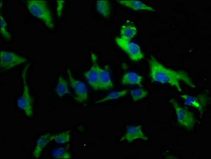
. TJP1 was detected in immunocytochemical section of A431 cells. Enzyme antigen retrieval was performed using IHC enzyme antigen retrieval reagent (AR0022) for 15 mins. The cells were blocked with 10% goat serum. And then incubated with 2microg/mL rabbit anti-TJP1 Antibody (PB9234) overnight at 4°C. DyLight?488 Conjugated Goat Anti-Rabbit IgG (BA1127) was used as secondary antibody at 1:100 dilution and incubated for 30 minutes at 37°C. The section was counterstained with DAPI. Visualize using a fluorescence microscope and filter sets appropriate for the label used.")
. Overlay histogram showing K562 cells stained with PB9234 (Blue line).The cells were blocked with 10% normal goat serum. And then incubated with rabbit anti-TJP1 Antibody (PB9234,1microg/1x106 cells) for 30 min at 20°C. DyLight?488 conjugated goat anti-rabbit IgG (BA1127, 5-10microg/1x106 cells) was used as secondary antibody for 30 minutes at 20°C. Isotype control antibody (Green line) was rabbit IgG (1microg/1x106) used under the same conditions. Unlabelled sample (Red line) was also used as a control.")
Figure 1. Western blot analysis of TJP1 using anti-TJP1 antibody (PB9234). Electrophoresis was performed on a 5-20% SDS-PAGE gel at 70V (Stacking gel) / 90V (Resolving gel) for 2-3 hours. The sample well of each lane was loaded with 30 ug of sample under reducing conditions. Lane 1: human PC-3 whole cell lysates, Lane 2: human CACO-2 whole cell lysates, Lane 3: human COLO320 whole cell lysates, Lane 4: rat testis tissue lysates, Lane 5: mouse testis tissue lysates. After electrophoresis, proteins were transferred to a nitrocellulose membrane at 150 mA for 50-90 minutes. Blocked the membrane with 5% non-fat milk/TBS for 1.5 hour at RT. The membrane was incubated with rabbit anti-TJP1 antigen affinity purified polyclonal antibody (Catalog # PB9234) at 0.5 microg/mL overnight at 4°C, then washed with TBS-0.1%Tween 3 times with 5 minutes each and probed with a goat anti-rabbit IgG-HRP secondary antibody at a dilution of 1:5000 for 1.5 hour at RT. The signal is developed using an Enhanced Chemiluminescent detection (ECL) kit (Catalog # EK1002) with Tanon 5200 system. A specific band was detected for TJP1 at approximately 220 kDa. The expected band size for TJP1 is at 185 kDa.
Anti-ZO1 tight junction protein/TJP1 Antibody Picoband(r)
PB9234-CARRIER-FREE
ApplicationsFlow Cytometry, ImmunoFluorescence, Western Blot, ImmunoCytoChemistry, ImmunoHistoChemistry
Product group Antibodies
ReactivityHuman, Mouse, Rat
TargetTJP1
Overview
- SupplierBoster Bio
- Product NameAnti-ZO1 tight junction protein/TJP1 Antibody Picoband(r)
- Delivery Days Customer9
- Application Supplier NoteWB: The detection limit for TJP1 is approximately 0.25ng/lane under reducing conditions. Tested Species: In-house tested species with positive results. By Heat: Boiling the paraffin sections in 10mM citrate buffer, pH6.0, for 20mins is required for the staining of formalin/paraffin sections. Other applications have not been tested. Optimal dilutions should be determined by end users.
- ApplicationsFlow Cytometry, ImmunoFluorescence, Western Blot, ImmunoCytoChemistry, ImmunoHistoChemistry
- CertificationResearch Use Only
- ClonalityPolyclonal
- Concentration500 ug/ml
- Gene ID7082
- Target nameTJP1
- Target descriptiontight junction protein 1
- Target synonymsZO-1, tight junction protein 1, tight junction protein ZO-1, zona occludens 1, zonula occludens 1 protein
- HostRabbit
- IsotypeIgG
- Protein IDQ07157
- Protein NameTight junction protein 1
- Scientific DescriptionBoster Bio Anti-ZO1 tight junction protein/TJP1 Antibody Picoband® catalog # PB9234. Tested in Flow Cytometry, IF, IHC, ICC, WB applications. This antibody reacts with Human, Mouse, Rat. The brand Picoband indicates this is a premium antibody that guarantees superior quality, high affinity, and strong signals with minimal background in Western blot applications. Only our best-performing antibodies are designated as Picoband, ensuring unmatched performance.
- ReactivityHuman, Mouse, Rat
- Storage Instruction-20°C,2°C to 8°C
- UNSPSC12352203
Related products
Product group Antibodies
TJP1 AntibodyCSB-PA023573HA01HU
ApplicationsImmunoFluorescence, ELISA
ReactivityHuman
TargetTJP1
- SizePrice
Product group Antibodies
Tjp1 Polyclonal AntibodyCAC11695
ApplicationsImmunoFluorescence, ELISA
TargetTJP1
- SizePrice
Product group Antibodies
Anti-ZO1 AntibodyA121581
ApplicationsImmunoFluorescence, Western Blot, ImmunoHistoChemistry, ImmunoHistoChemistry Frozen, ImmunoHistoChemistry Paraffin
ReactivityCanine, Human, Monkey, Mouse, Rat
- SizePrice
Product group Antibodies
Anti-ZO-1 Antibody144-61634
ApplicationsImmunoFluorescence, Western Blot
ReactivityHuman, Mouse, Rat
TargetTJP1
- SizePrice
Product group Antibodies

TJP1 / ZO-1 AntibodyLS-C831674
ApplicationsELISA, ImmunoHistoChemistry
ReactivityHuman, Mouse, Rat
TargetTJP1
- SizePrice
Product group Antibodies

Goat anti-ZO-1EB09206
ApplicationsImmunoFluorescence, ELISA
ReactivityHuman
TargetTJP1
- SizePrice

Product group Antibodies
Anti-TJP1 AntibodyHPA001636
ApplicationsWestern Blot, ImmunoCytoChemistry, ImmunoHistoChemistry
ReactivityHuman
TargetTJP1
- SizePrice
![Non-transfected (–) and transfected (+) HeLa whole cell extracts (30 μg) were separated by 5% SDS-PAGE, and the membrane was blotted with ZO-1 antibody [C3], C-term (GTX108587) diluted at 1:500.](https://www.genetex.com/upload/website/prouct_img/normal/GTX108587/GTX108587_40618_20160804_WB_shRNA_watermark_w_23060120_565.webp)
Product group Antibodies
ZO-1 antibody [C3], C-termGTX108587
ApplicationsWestern Blot
ReactivityHuman
TargetTJP1
- SizePrice