

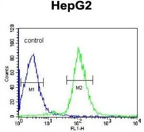
FACS analysis of HepG2 cells using GTX81758 Antithrombin III antibody, C-term. Green : primary antibody Blue : negative control

 using GTX81758 Antithrombin III antibody, C-term.")


FACS analysis of HepG2 cells using GTX81758 Antithrombin III antibody, C-term. Green : primary antibody Blue : negative control
Antithrombin III antibody, C-term
GTX81758
ApplicationsFlow Cytometry, ImmunoFluorescence, Western Blot, ImmunoCytoChemistry, ImmunoHistoChemistry, ImmunoHistoChemistry Paraffin
Product group Antibodies
ReactivityHuman, Mouse
TargetSERPINC1
Overview
- SupplierGeneTex
- Product NameAntithrombin III antibody, C-term
- Delivery Days Customer9
- Application Supplier NoteWB: 1:1000. ICC/IF: 1:10-1:50. IHC-P: 1:50-1:100. FCM: 1:10-1:50. *Optimal dilutions/concentrations should be determined by the researcher.Not tested in other applications.
- ApplicationsFlow Cytometry, ImmunoFluorescence, Western Blot, ImmunoCytoChemistry, ImmunoHistoChemistry, ImmunoHistoChemistry Paraffin
- CertificationResearch Use Only
- ClonalityPolyclonal
- ConjugateUnconjugated
- Gene ID462
- Target nameSERPINC1
- Target descriptionserpin family C member 1
- Target synonymsAT3, AT3D, ATIII, ATIII-R2, ATIII-T1, ATIII-T2, THPH7, antithrombin-III, serine (or cysteine) proteinase inhibitor, clade C (antithrombin), member 1, serpin peptidase inhibitor clade C member 1, serpin peptidase inhibitor, clade C (antithrombin), member 1
- HostRabbit
- IsotypeIgG
- Protein IDP01008
- Protein NameAntithrombin-III
- Scientific DescriptionThe protein encoded by this gene is a plasma protease inhibitor and a member of the serpin superfamily. This protein inhibits thrombin as well as other activated serine proteases of the coagulation system, and it regulates the blood coagulation cascade. The protein includes two functional domains: the heparin binding-domain at the N-terminus of the mature protein, and the reactive site domain at the C-terminus. The inhibitory activity is enhanced by the presence of heparin. More than 120 mutations have been identified for this gene, many of which are known to cause antithrombin-III deficiency. [provided by RefSeq, Jul 2009]
- ReactivityHuman, Mouse
- Storage Instruction-20°C or -80°C,2°C to 8°C
- UNSPSC41116161
Datasheet
Related products
Product group Antibodies
Anti-SERPINC1 AntibodyA28836
ApplicationsWestern Blot
ReactivityHuman, Mouse, Rat
- SizePrice
Product group Antibodies
Anti-SERPINC1 Antibody144-01574
ApplicationsImmunoFluorescence, Western Blot, ImmunoHistoChemistry
ReactivityHuman, Mouse
TargetSERPINC1
- SizePrice

Product group Antibodies
Anti-Antithrombin III/SERPINC1 Antibody Picoband(r)A00469-1-CARRIER-FREE
ApplicationsFlow Cytometry, Western Blot, ELISA
ReactivityHuman, Mouse, Rat
TargetSERPINC1
- SizePrice
Product group Antibodies
SERPINC1 Recombinant Antibody, Biotin ConjugatedBSM-61553R-BIOTIN
ApplicationsWestern Blot
ReactivityHuman
TargetSERPINC1
- SizePrice
Product group Antibodies
SERPINC1 Polyclonal AntibodyCAC14879
ApplicationsWestern Blot, ELISA, ImmunoHistoChemistry
ReactivityRat
TargetSERPINC1
- SizePrice
Product group Antibodies
SERPINC1 AntibodyCSB-PA021079LA01HU
ApplicationsWestern Blot, ELISA, ImmunoHistoChemistry
ReactivityHuman, Rat
TargetSERPINC1
- SizePrice
Product group Antibodies
Antithrombin III antibodyGTX30705
ApplicationsELISA
ReactivityHuman
TargetSERPINC1
- SizePrice

Product group Antibodies

Antithrombin-III AntibodyLS-C331551
ApplicationsImmunoFluorescence, Western Blot, ImmunoHistoChemistry
ReactivityHuman, Mouse
TargetSERPINC1
- SizePrice