

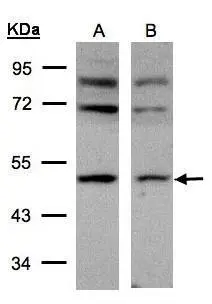
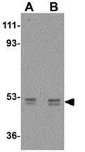
Sample(30 μg of whole cell lysate) A:293T B:HeLa S3(GTX14654) 10% SDS PAGE GTX101502 diluted at 1:500
 antibody at 1:100 dilution.
Antigen Retrieval: Trilogy? (EDTA based, pH 8.0) buffer, 15min")
Sample(30 μg of whole cell lysate) A:293T B:HeLa S3(GTX14654) 10% SDS PAGE GTX101502 diluted at 1:500
AP2M1 antibody
GTX101502
ApplicationsWestern Blot, ImmunoHistoChemistry, ImmunoHistoChemistry Paraffin
Product group Antibodies
ReactivityHuman
TargetAP2M1
Overview
- SupplierGeneTex
- Product NameAP2M1 antibody
- Delivery Days Customer9
- Application Supplier NoteWB: 1:500-1:3000. IHC-P: 1:100-1:1000. *Optimal dilutions/concentrations should be determined by the researcher.Not tested in other applications.
- ApplicationsWestern Blot, ImmunoHistoChemistry, ImmunoHistoChemistry Paraffin
- CertificationResearch Use Only
- ClonalityPolyclonal
- Concentration0.57 mg/ml
- ConjugateUnconjugated
- Gene ID1173
- Target nameAP2M1
- Target descriptionadaptor related protein complex 2 subunit mu 1
- Target synonymsAP50, CLAPM1, MRD60, mu2, AP-2 complex subunit mu, AP-2 mu 2 chain, HA2 50 kDA subunit, adaptin-mu2, adaptor protein complex AP-2 subunit mu, adaptor related protein complex 2 mu 1 subunit, adaptor-related protein complex 2 subunit mu, clathrin adaptor complex AP2, mu subunit, clathrin assembly protein complex 2 medium chain, clathrin assembly protein complex 2 mu medium chain, clathrin coat adaptor protein AP50, clathrin coat assembly protein AP50, clathrin coat-associated protein AP50, clathrin-associated/assembly/adaptor protein, medium 1, plasma membrane adaptor AP-2 50 kDa protein, plasma membrane adaptor AP-2 50kDA protein
- HostRabbit
- IsotypeIgG
- Protein IDQ96CW1
- Protein NameAP-2 complex subunit mu
- Scientific DescriptionThis gene encodes a subunit of the heterotetrameric coat assembly protein complex 2 (AP2), which belongs to the adaptor complexes medium subunits family. The encoded protein is required for the activity of a vacuolar ATPase, which is responsible for proton pumping occurring in the acidification of endosomes and lysosomes. The encoded protein may also play an important role in regulating the intracellular trafficking and function of CTLA-4 protein. Two transcript variants encoding different isoforms have been found for this gene. [provided by RefSeq]
- ReactivityHuman
- Storage Instruction-20°C or -80°C,2°C to 8°C
- UNSPSC41116161
Datasheet
Related products
Product group Antibodies
Anti-AP2M1 AntibodyA28295
ApplicationsWestern Blot
ReactivityHuman, Mouse, Rat
- SizePrice

Product group Antibodies
Anti-AP2M1 Antibody Picoband(r)A06179-3-CARRIER-FREE
ApplicationsFlow Cytometry, ImmunoFluorescence, Western Blot, ELISA, ImmunoCytoChemistry
ReactivityHuman, Mouse, Rat
TargetAP2M1
- SizePrice
Product group Antibodies
Anti-AP2M1 Antibody144-02492
ApplicationsWestern Blot, ImmunoHistoChemistry
ReactivityHuman, Mouse, Rat
TargetAP2M1
- SizePrice
Product group Antibodies

AP50 / AP2M1 AntibodyLS-C748977
ApplicationsImmunoFluorescence
ReactivityHuman
TargetAP2M1
- SizePrice
Product group Antibodies
AP2M1 Recombinant Antibody, Biotin ConjugatedBSM-61305R-BIOTIN
ApplicationsWestern Blot
TargetAP2M1
- SizePrice
Product group Antibodies
AP2M1 AntibodyCSB-PA00625A0RB
ApplicationsWestern Blot, ELISA, ImmunoHistoChemistry
ReactivityHuman
TargetAP2M1
- SizePrice
Product group Antibodies
Ap2M1 Polyclonal AntibodyCAC07420
ApplicationsWestern Blot, ELISA, ImmunoHistoChemistry
TargetAP2M1
- SizePrice
Product group Antibodies
AP2M1 antibodyGTX30052
ApplicationsWestern Blot
ReactivityHuman
TargetAP2M1
- SizePrice
Product group Antibodies
AP2M1 antibodyGTX85110
ApplicationsWestern Blot, ELISA, ImmunoHistoChemistry, ImmunoHistoChemistry Paraffin
ReactivityHuman, Mouse, Rat
TargetAP2M1
- SizePrice