

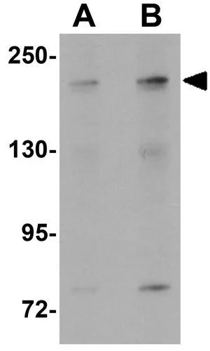
WB analysis of SK-N-SH cell lysate using GTX31801 APC1 antibody. Working concentration : (A) 1 and (B) 2 μg/ml

WB analysis of SK-N-SH cell lysate using GTX31801 APC1 antibody. Working concentration : (A) 1 and (B) 2 μg/ml
APC1 antibody
GTX31801
ApplicationsWestern Blot, ELISA, ImmunoHistoChemistry, ImmunoHistoChemistry Paraffin
Product group Antibodies
ReactivityHuman, Mouse
TargetANAPC1
Overview
- SupplierGeneTex
- Product NameAPC1 antibody
- Delivery Days Customer9
- Application Supplier NoteWB: 1 - 2 microg/mL. IHC-P: 5 microg/mL. *Optimal dilutions/concentrations should be determined by the researcher.Not tested in other applications.
- ApplicationsWestern Blot, ELISA, ImmunoHistoChemistry, ImmunoHistoChemistry Paraffin
- CertificationResearch Use Only
- ClonalityPolyclonal
- Concentration1 mg/ml
- ConjugateUnconjugated
- Gene ID64682
- Target nameANAPC1
- Target descriptionanaphase promoting complex subunit 1
- Target synonymsAPC1, MCPR, TSG24, anaphase-promoting complex subunit 1, anaphase-promoting complex 1 (meiotic checkpoint regulator), cyclosome subunit 1, mitotic checkpoint regulator, testis-specific gene 24 protein
- HostRabbit
- IsotypeIgG
- Protein IDQ9H1A4
- Protein NameAnaphase-promoting complex subunit 1
- Scientific DescriptionThis gene encodes a subunit of the anaphase-promoting complex. This complex is an E3 ubiquitin ligase that regulates progression through the metaphase to anaphase portion of the cell cycle by ubiquitinating proteins which targets them for degradation. [provided by RefSeq, Dec 2011]
- ReactivityHuman, Mouse
- Storage Instruction-20°C or -80°C,2°C to 8°C
- UNSPSC41116161
Datasheet
Related products
Product group Antibodies
ANAPC1 AntibodyCSB-PA000898
ApplicationsImmunoFluorescence, Western Blot, ELISA
ReactivityHuman, Mouse
TargetANAPC1
- SizePrice

Product group Antibodies
Anti-APC1/ANAPC1 Antibody Picoband(r)A03471-2-CARRIER-FREE
ApplicationsFlow Cytometry, ImmunoFluorescence, Western Blot, ELISA, ImmunoCytoChemistry, ImmunoHistoChemistry
ReactivityHuman
TargetANAPC1
- SizePrice
Product group Antibodies
Anti-ANAPC1 AntibodyA29366
ApplicationsWestern Blot
ReactivityHuman, Mouse, Rat
- SizePrice

Product group Antibodies
Anti-ANAPC1 AntibodyHPA036329
ApplicationsWestern Blot, ImmunoHistoChemistry
ReactivityHuman
TargetANAPC1
- SizePrice
Product group Antibodies

ANAPC1 / APC1 AntibodyLS-C332600
ApplicationsWestern Blot
ReactivityHuman
TargetANAPC1
- SizePrice
![Western blot using GeneTex Affinity Purified anti-APC1 pS355 antibody (GTX10923) shows detection of a band ~215 kDa corresponding to phosphorylated human APC1 (arrowhead). Lane 1 shows lysate from asynchronous cells. Lane 2 shows lysate from cells treated with thymidine to synchronize cells at the G1/S boundary. Lane 3 shows lysate from cells treated with nocodazole to synchronize cells at the M phase. Phosphorylated APC1 is mostly present only in cell preparations arrested at cell division. Each lane contains approximately 30 ug of HeLa S3 whole cell lysates separated by 12.5% SDS-PAGE followed by transfer to nitrocellulose. After blocking with 5% non-fat dry milk in TTBS, the membrane was probed with the primary antibody diluted to 1:500 for 1 h at room temperature followed by washes and reaction with a 1:5,000 dilution of HRP Gt-a-Rabbit IgG [H&L] MX (GTX27090) for 45 min at room temperature. ECL reagent was used for detection. Other detection systems will yield similar results.](https://www.genetex.com/upload/website/prouct_img/normal/GTX10923/GTX10923_20160330_WB_w_23060120_216.webp)
Product group Antibodies
APC1 (phospho Ser355) antibodyGTX10923
ApplicationsImmunoPrecipitation, Western Blot, ELISA
ReactivityHuman
TargetANAPC1
- SizePrice
Product group Antibodies
ApplicationsImmunoFluorescence, ELISA, ImmunoCytoChemistry, ImmunoHistoChemistry, ImmunoHistoChemistry Frozen, ImmunoHistoChemistry Paraffin
ReactivityChicken, Human, Mouse, Rat
TargetANAPC1
- SizePrice