

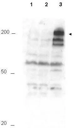
Western blot using GeneTex Affinity Purified anti-APC1 pS355 antibody (GTX10923) shows detection of a band ~215 kDa corresponding to phosphorylated human APC1 (arrowhead). Lane 1 shows lysate from asynchronous cells. Lane 2 shows lysate from cells treated with thymidine to synchronize cells at the G1/S boundary. Lane 3 shows lysate from cells treated with nocodazole to synchronize cells at the M phase. Phosphorylated APC1 is mostly present only in cell preparations arrested at cell division. Each lane contains approximately 30 ug of HeLa S3 whole cell lysates separated by 12.5% SDS-PAGE followed by transfer to nitrocellulose. After blocking with 5% non-fat dry milk in TTBS, the membrane was probed with the primary antibody diluted to 1:500 for 1 h at room temperature followed by washes and reaction with a 1:5,000 dilution of HRP Gt-a-Rabbit IgG [H&L] MX (GTX27090) for 45 min at room temperature. ECL reagent was used for detection. Other detection systems will yield similar results.
Western blot using GeneTex Affinity Purified anti-APC1 pS355 antibody (GTX10923) shows detection of a band ~215 kDa corresponding to phosphorylated human APC1 (arrowhead). Lane 1 shows lysate from asynchronous cells. Lane 2 shows lysate from cells treated with thymidine to synchronize cells at the G1/S boundary. Lane 3 shows lysate from cells treated with nocodazole to synchronize cells at the M phase. Phosphorylated APC1 is mostly present only in cell preparations arrested at cell division. Each lane contains approximately 30 ug of HeLa S3 whole cell lysates separated by 12.5% SDS-PAGE followed by transfer to nitrocellulose. After blocking with 5% non-fat dry milk in TTBS, the membrane was probed with the primary antibody diluted to 1:500 for 1 h at room temperature followed by washes and reaction with a 1:5,000 dilution of HRP Gt-a-Rabbit IgG [H&L] MX (GTX27090) for 45 min at room temperature. ECL reagent was used for detection. Other detection systems will yield similar results.
APC1 (phospho Ser355) antibody
GTX10923
ApplicationsImmunoPrecipitation, Western Blot, ELISA
Product group Antibodies
ReactivityHuman
TargetANAPC1
Overview
- SupplierGeneTex
- Product NameAPC1 (phospho Ser355) antibody
- Delivery Days Customer9
- Application Supplier NoteWB: 1:200-1:2000. IP: 1:100. ELISA: 1:10000-1:35000. *Optimal dilutions/concentrations should be determined by the researcher.Not tested in other applications.
- ApplicationsImmunoPrecipitation, Western Blot, ELISA
- CertificationResearch Use Only
- ClonalityPolyclonal
- Concentration1 mg/ml
- ConjugateUnconjugated
- Gene ID64682
- Target nameANAPC1
- Target descriptionanaphase promoting complex subunit 1
- Target synonymsAPC1, MCPR, TSG24, anaphase-promoting complex subunit 1, anaphase-promoting complex 1 (meiotic checkpoint regulator), cyclosome subunit 1, mitotic checkpoint regulator, testis-specific gene 24 protein
- HostRabbit
- IsotypeIgG
- Protein IDQ9H1A4
- Protein NameAnaphase-promoting complex subunit 1
- Scientific DescriptionThis is one of a number of novel phosphorylation sites for APC1 identified in Kraft C et al. It may be involved in the regulation of the APC/C.
- ReactivityHuman
- Storage Instruction-20°C or -80°C,2°C to 8°C
- UNSPSC41116161
Datasheet
Related products
Product group Antibodies
Anti-ANAPC1 AntibodyA29366
ApplicationsWestern Blot
ReactivityHuman, Mouse, Rat
- SizePrice

Product group Antibodies
Anti-APC1/ANAPC1 Antibody Picoband(r)A03471-2-CARRIER-FREE
ApplicationsFlow Cytometry, ImmunoFluorescence, Western Blot, ELISA, ImmunoCytoChemistry, ImmunoHistoChemistry
ReactivityHuman
TargetANAPC1
- SizePrice
Product group Antibodies
ApplicationsImmunoFluorescence, ELISA, ImmunoCytoChemistry, ImmunoHistoChemistry, ImmunoHistoChemistry Frozen, ImmunoHistoChemistry Paraffin
ReactivityChicken, Human, Mouse, Rat
TargetANAPC1
- SizePrice
Product group Antibodies
ANAPC1 AntibodyCSB-PA000898
ApplicationsImmunoFluorescence, Western Blot, ELISA
ReactivityHuman, Mouse
TargetANAPC1
- SizePrice
Product group Antibodies
APC1 antibodyGTX31801
ApplicationsWestern Blot, ELISA, ImmunoHistoChemistry, ImmunoHistoChemistry Paraffin
ReactivityHuman, Mouse
TargetANAPC1
- SizePrice
Product group Antibodies

ANAPC1 / APC1 AntibodyLS-C332600
ApplicationsWestern Blot
ReactivityHuman
TargetANAPC1
- SizePrice

Product group Antibodies
Anti-ANAPC1 AntibodyHPA036329
ApplicationsWestern Blot, ImmunoHistoChemistry
ReactivityHuman
TargetANAPC1
- SizePrice