

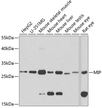
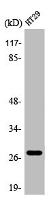
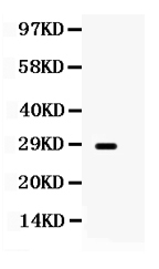
WB analysis of various sample lysates using GTX54297 Aquaporin 0 antibody. Dilution : 1:1000 Loading : 25μg per lane


WB analysis of various sample lysates using GTX54297 Aquaporin 0 antibody. Dilution : 1:1000 Loading : 25μg per lane
Aquaporin 0 antibody
GTX54297
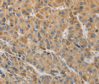
ApplicationsWestern Blot, ImmunoHistoChemistry, ImmunoHistoChemistry Paraffin
Product group Antibodies
ReactivityHuman, Mouse, Rat
TargetMIP
Overview
- SupplierGeneTex
- Product NameAquaporin 0 antibody
- Delivery Days Customer7
- Application Supplier NoteWB: 1:500 - 1:1000. IHC-P: 1:100 - 1:200. *Optimal dilutions/concentrations should be determined by the researcher.Not tested in other applications.
- ApplicationsWestern Blot, ImmunoHistoChemistry, ImmunoHistoChemistry Paraffin
- CertificationResearch Use Only
- ClonalityPolyclonal
- ConjugateUnconjugated
- Gene ID4284
- Target nameMIP
- Target descriptionmajor intrinsic protein of lens fiber
- Target synonymsAQP0, CTRCT15, LIM1, MIP26, MP26, lens fiber major intrinsic protein, aquaporin 0
- HostRabbit
- IsotypeIgG
- Protein IDP30301
- Protein NameLens fiber major intrinsic protein
- Scientific DescriptionMajor intrinsic protein is a member of the water-transporting aquaporins as well as the original member of the MIP family of channel proteins. The function of the fiber cell membrane protein encoded by this gene is undetermined, yet this protein is speculated to play a role in intracellular communication. The MIP protein is expressed in the ocular lens and is required for correct lens function. This gene has been mapped among aquaporins AQP2, AQP5, and AQP6, in a potential gene cluster at 12q13. [provided by RefSeq, Jul 2008]
- ReactivityHuman, Mouse, Rat
- Storage Instruction-20°C or -80°C,2°C to 8°C
- UNSPSC41116161
Datasheet
Related products
Product group Antibodies
Anti-MIP AntibodyA47194
ApplicationsImmunoHistoChemistry
ReactivityHuman
- SizePrice
Product group Antibodies
Anti-MIP Antibody144-02886
ApplicationsWestern Blot, ImmunoHistoChemistry
ReactivityHuman, Mouse, Rat
TargetMIP
- SizePrice
Product group Antibodies

MIP / AQP0 / Aquaporin 0 AntibodyLS-C768160
ApplicationsELISA, ImmunoHistoChemistry
ReactivityHuman, Mouse, Rat
TargetMIP
- SizePrice
Product group Antibodies
AQP0 Polyclonal AntibodyBS-6661R
ApplicationsImmunoFluorescence, ELISA, ImmunoCytoChemistry, ImmunoHistoChemistry, ImmunoHistoChemistry Frozen, ImmunoHistoChemistry Paraffin
ReactivityBovine, Chicken, Equine, Guinea Pig, Human, Mouse, Porcine, Rabbit, Rat, Sheep
TargetMIP
- SizePrice
Product group Antibodies
MIP AntibodyCSB-PA000910
ApplicationsWestern Blot, ELISA, ImmunoHistoChemistry
ReactivityHuman, Mouse, Rat
TargetMIP
- SizePrice

Product group Antibodies
Anti-MIP AntibodyHPA014940
ApplicationsImmunoHistoChemistry
ReactivityHuman
TargetMIP
- SizePrice
Product group Antibodies
Anti-MIP AntibodyCAB2886
ApplicationsWestern Blot, ELISA, ImmunoHistoChemistry, ImmunoHistoChemistry Paraffin
ReactivityHuman
TargetMIP
- SizePrice
Product group Antibodies
Anti-Aquaporin 0/MIP Antibody Picoband(r)PB9811-CARRIER-FREE
ApplicationsWestern Blot
ReactivityHuman, Rat
TargetMIP
- SizePrice