

MIP antibody
10R-10490
Product group Antibodies
Overview
- SupplierBiosynth
- Product NameMIP antibody
- Delivery Days Customer11
- CertificationResearch Use Only
- UNSPSC41116161
Related products
Product group Antibodies
Anti-MIP AntibodyA47194
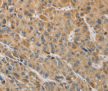
ApplicationsImmunoHistoChemistry
ReactivityHuman
- SizePrice
Product group Antibodies
Anti-MIP Antibody144-02886
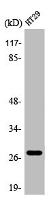
ApplicationsWestern Blot, ImmunoHistoChemistry
ReactivityHuman, Mouse, Rat
TargetMIP
- SizePrice
Product group Antibodies

MIP / AQP0 / Aquaporin 0 AntibodyLS-C768160
ApplicationsELISA, ImmunoHistoChemistry
ReactivityHuman, Mouse, Rat
TargetMIP
- SizePrice
Product group Antibodies
AQP0 Polyclonal AntibodyBS-6661R
ApplicationsImmunoFluorescence, ELISA, ImmunoCytoChemistry, ImmunoHistoChemistry, ImmunoHistoChemistry Frozen, ImmunoHistoChemistry Paraffin
ReactivityBovine, Chicken, Equine, Guinea Pig, Human, Mouse, Porcine, Rabbit, Rat, Sheep
TargetMIP
- SizePrice
Product group Antibodies
MIP AntibodyCSB-PA000910
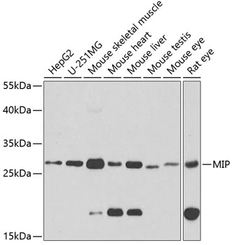
ApplicationsWestern Blot, ELISA, ImmunoHistoChemistry
ReactivityHuman, Mouse, Rat
TargetMIP
- SizePrice
Product group Antibodies
Aquaporin 0 antibodyGTX54297
ApplicationsWestern Blot, ImmunoHistoChemistry, ImmunoHistoChemistry Paraffin
ReactivityHuman, Mouse, Rat
TargetMIP
- SizePrice

Product group Antibodies
Anti-MIP AntibodyHPA014940
ApplicationsImmunoHistoChemistry
ReactivityHuman
TargetMIP
- SizePrice
Product group Antibodies
Anti-Aquaporin 0/MIP Antibody Picoband(r)PB9811-CARRIER-FREE
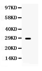
ApplicationsWestern Blot
ReactivityHuman, Rat
TargetMIP
- SizePrice
Product group Antibodies
Anti-MIP AntibodyCAB2886
ApplicationsWestern Blot, ELISA, ImmunoHistoChemistry, ImmunoHistoChemistry Paraffin
ReactivityHuman
TargetMIP
- SizePrice