

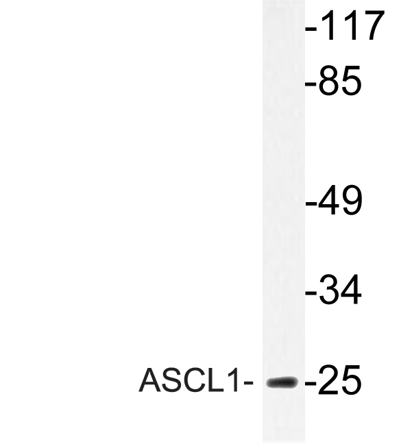
ASCL1 Polyclonal Antibody
CAC15005
ApplicationsWestern Blot, ELISA, ImmunoHistoChemistry
Product group Antibodies
ReactivityRat
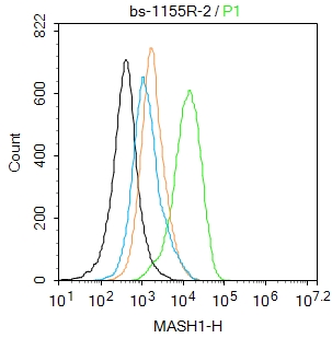
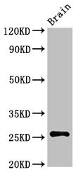
TargetASCL1
Overview
- SupplierBiomatik
- Product NameASCL1 Polyclonal Antibody
- Delivery Days Customer12
- ApplicationsWestern Blot, ELISA, ImmunoHistoChemistry
- CertificationResearch Use Only
- ClonalityPolyclonal
- ConjugateUnconjugated
- Gene ID429
- Target nameASCL1
- Target descriptionachaete-scute family bHLH transcription factor 1
- Target synonymsASH1, HASH1, MASH1, bHLHa46, achaete-scute homolog 1, ASH-1, achaete scute protein, achaete-scute complex homolog 1, achaete-scute complex-like 1, class A basic helix-loop-helix protein 46
- HostRabbit
- IsotypeIgG
- Protein IDP50553
- Protein NameAchaete-scute homolog 1
- Scientific DescriptionThe ASCL1 Polyclonal Antibody (Species: Human) has been validated for the following applications: ELISA, WB, IHC.
- ReactivityRat
- Storage Instruction-20°C,2°C to 8°C
- UNSPSC12352203
Related products
Product group Antibodies
Anti-ASCL1 AntibodyA95934
ApplicationsWestern Blot, ELISA, ImmunoHistoChemistry
ReactivityHuman, Mouse, Rat
- SizePrice
Product group Antibodies

Anti-ASCL1 [SC72.2]Ab02398-1.1
ApplicationsELISA, ImmunoHistoChemistry
ReactivityHuman
TargetASCL1
- SizePrice
Product group Antibodies
ApplicationsWestern Blot
TargetASCL1
- SizePrice

Product group Antibodies
Anti-ASCL1 Antibody Picoband(r)A03023-2-CARRIER-FREE
ApplicationsFlow Cytometry, Western Blot, ImmunoHistoChemistry
ReactivityHuman, Mouse, Rat
TargetASCL1
- SizePrice
Product group Antibodies
References
ASCL1 Polyclonal AntibodyBS-1155R
ApplicationsImmunoFluorescence, Western Blot, ELISA, ImmunoCytoChemistry, ImmunoHistoChemistry, ImmunoHistoChemistry Frozen, ImmunoHistoChemistry Paraffin
ReactivityBovine, Human, Mouse, Rat, Sheep
TargetASCL1
- SizePrice
Product group Antibodies
ASCL1 AntibodyCSB-PA002199LA01HU
ApplicationsWestern Blot, ELISA, ImmunoHistoChemistry
ReactivityHuman, Rat
TargetASCL1
- SizePrice
Product group Antibodies

Goat anti-ASCL1 (aa79-91)EB12072
ApplicationsImmunoFluorescence, Western Blot, ELISA
ReactivityBovine, Canine, Human, Mouse, Rat
TargetASCL1
- SizePrice
Product group Antibodies

ASCL1 / MASH1 AntibodyLS-C402152
ApplicationsELISA, ImmunoHistoChemistry
ReactivityHuman, Mouse, Rat
TargetASCL1
- SizePrice

Product group Antibodies
Anti-ASCL1 AntibodyHPA076307
ApplicationsImmunoCytoChemistry
ReactivityHuman
TargetASCL1
- SizePrice