

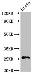
Western Blot Positive WB detected in: Rat brain tissue All lanes: ASCL1 antibody at 3ug/ml Secondary Goat polyclonal to rabbit IgG at 1/50000 dilution Predicted band size: 26 kDa Observed band size: 26 kDa

Western Blot Positive WB detected in: Rat brain tissue All lanes: ASCL1 antibody at 3ug/ml Secondary Goat polyclonal to rabbit IgG at 1/50000 dilution Predicted band size: 26 kDa Observed band size: 26 kDa
ASCL1 Antibody
CSB-PA002199LA01HU
ApplicationsWestern Blot, ELISA, ImmunoHistoChemistry
Product group Antibodies
ReactivityHuman, Rat
TargetASCL1
Overview
- SupplierCusabio
- Product NameASCL1 Antibody
- Delivery Days Customer20
- ApplicationsWestern Blot, ELISA, ImmunoHistoChemistry
- CertificationResearch Use Only
- ClonalityPolyclonal
- ConjugateUnconjugated
- Gene ID429
- Target nameASCL1
- Target descriptionachaete-scute family bHLH transcription factor 1
- Target synonymsASH1, HASH1, MASH1, bHLHa46, achaete-scute homolog 1, ASH-1, achaete scute protein, achaete-scute complex homolog 1, achaete-scute complex-like 1, class A basic helix-loop-helix protein 46
- HostRabbit
- IsotypeIgG
- Protein IDP50553
- Protein NameAchaete-scute homolog 1
- Scientific DescriptionTranscription factor that controls transcriptional expression of its target genes by binding to the E box (5-CANNTG-3). Plays a role at early stages of development of specific neural lineages in most regions of the CNS, and of several lineages in the PNS. Acts synergistically with FOXN4 to specify the identity of V2b neurons rather than V2a from bipotential p2 progenitors during spinal cord neurogenesis, probably through DLL4-NOTCH signaling activation. Essential for the generation of olfactory and autonomic neurons (By similarity).
- ReactivityHuman, Rat
- Storage Instruction-20°C or -80°C
- UNSPSC41116161
Related products
Product group Antibodies
Anti-ASCL1 AntibodyA95934
ApplicationsWestern Blot, ELISA, ImmunoHistoChemistry
ReactivityHuman, Mouse, Rat
- SizePrice
Product group Antibodies

Anti-ASCL1 [SC72.2]Ab02398-1.1
ApplicationsELISA, ImmunoHistoChemistry
ReactivityHuman
TargetASCL1
- SizePrice
Product group Antibodies
ApplicationsWestern Blot
TargetASCL1
- SizePrice

Product group Antibodies
Anti-ASCL1 Antibody Picoband(r)A03023-2-CARRIER-FREE
ApplicationsFlow Cytometry, Western Blot, ImmunoHistoChemistry
ReactivityHuman, Mouse, Rat
TargetASCL1
- SizePrice
Product group Antibodies
References
ASCL1 Polyclonal AntibodyBS-1155R
ApplicationsImmunoFluorescence, Western Blot, ELISA, ImmunoCytoChemistry, ImmunoHistoChemistry, ImmunoHistoChemistry Frozen, ImmunoHistoChemistry Paraffin
ReactivityBovine, Human, Mouse, Rat, Sheep
TargetASCL1
- SizePrice
Product group Antibodies

Goat anti-ASCL1 (aa79-91)EB12072
ApplicationsImmunoFluorescence, Western Blot, ELISA
ReactivityBovine, Canine, Human, Mouse, Rat
TargetASCL1
- SizePrice
Product group Antibodies
ASCL1 Polyclonal AntibodyCAC15005
ApplicationsWestern Blot, ELISA, ImmunoHistoChemistry
ReactivityRat
TargetASCL1
- SizePrice
Product group Antibodies

ASCL1 / MASH1 AntibodyLS-C402152
ApplicationsELISA, ImmunoHistoChemistry
ReactivityHuman, Mouse, Rat
TargetASCL1
- SizePrice

Product group Antibodies
Anti-ASCL1 AntibodyHPA076307
ApplicationsImmunoCytoChemistry
ReactivityHuman
TargetASCL1
- SizePrice